
Page 82 - AACN Essentials of Critical-Care Nursing Pocket Handbook, Second Edition
P. 82
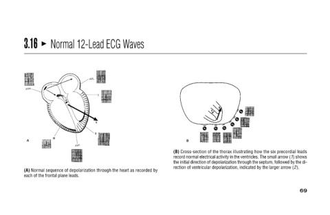
record normal electrical activity in the ventricles. The small arrow (1) shows
                                                   the initial direction of depolarization through the septum, followed by the di-
                                                            69
                                                (B) Cross-section of the thorax illustrating how the six precordial leads
                                                     rection of ventricular depolarization, indicated by the larger arrow (2).


                Normal 12-Lead ECG Waves              (A) Normal sequence of depolarization through the heart as recorded by


                                                       each of the frontal plane leads.
                3.16