
Page 106 - untitled
P. 106
AAAC46 21/5/05 10:53 AM Page 105
Iliolumbar
Arteries from capsule in retinacula
ligament
Body weight
Artery in
ligamentum teres
Long and short
posterior ligaments
Greater sciatic
foramen
Sacrospinous
ligament Sacrotuberous
Sacrotuberous ligament
ligament
Subcapital
Fig.46.5 Ischiofemoral ligament Cervical Intracapsular
Basal
The ligaments of the back of the hip.
Pertrochanteric Extracapsular
The smaller diagram shows how the sacrotuberous and
sacrospinous ligaments resist rotation of the sacrum
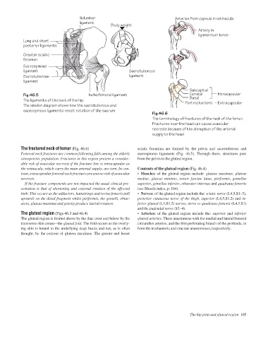
Fig.46.6
The terminology of fractures of the neck of the femur.
Fractures near the head can cause avascular
necrosis because of the disruption of the arterial
supply to the head
The fractured neck of femur (Fig. 46.6) sciatic foramina are formed by the pelvis and sacrotuberous and
Femoral neck fractures are common following falls among the elderly sacrospinous ligaments (Fig. 46.5). Through these, structures pass
osteoporotic population. Fractures in this region present a consider- from the pelvis to the gluteal region.
able risk of avascular necrosis if the fracture line is intracapsular as
the retinacula, which carry the main arterial supply, are torn. In con- Contents of the gluteal region (Fig. 46.4)
trast, extracapsular femoral neck fractures present no risk of avascular • Muscles: of the gluteal region include: gluteus maximus, gluteus
necrosis. medius, gluteus minimis, tensor fasciae latae, piriformis, gemellus
If the fracture components are not impacted the usual clinical pre- superior, gemellus inferior, obturator internus and quadratus femoris
sentation is that of shortening and external rotation of the affected (see Muscle index, p. 164).
limb. This occurs as the adductors, hamstrings and rectus femoris pull • Nerves: of the gluteal region include the: sciatic nerve (L4,5,S1–3),
upwards on the distal fragment whilst piriformis, the gemelli, obtur- posterior cutaneous nerve of the thigh, superior (L4,5,S1,2) and in-
ators, gluteus maximus and gravity produce lateral rotation. ferior gluteal (L5,S1,2) nerves, nerve to quadratus femoris (L4,5,S1)
and the pudendal nerve (S2–4).
The gluteal region (Figs 46.3 and 46.4) • Arteries: of the gluteal region include the: superior and inferior
The gluteal region is limited above by the iliac crest and below by the gluteal arteries. These anastomose with the medial and lateral femoral
transverse skin creaseathe gluteal fold. The fold occurs as the overly- circumflex arteries, and the first perforating branch of the profunda, to
ing skin is bound to the underlying deep fascia and not, as is often form the trochanteric and cruciate anastomoses, respectively.
thought, by the contour of gluteus maximus. The greater and lesser
The hip joint and gluteal region 105