
Page 91 - untitled
P. 91
AAAC40 21/5/05 10:47 AM Page 90
40 Surface anatomy of the upper limb
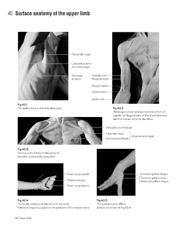
Pectoralis major
Latissimus dorsi
and teres major
Serratus Cephalic vein
anterior Biceps brachii
Biceps tendon
Aponeurosis
Basilic vein
Fig.40.1
The axilla with the arm fully abducted Fig.40.2
The biceps tendon and aponeurosis which are
a guide to the positions of the brachial artery
and the median nerve at the elbow
Deltopectoral triangle
Clavicular head
of pectoralis major
Sternocostal head
Fig.40.3
Strong contraction of the pectoral
muscles produced by adduction
Flexor carpi radialis Extensor pollicis longus
Extensor pollicis brevis
Palmaris longus
Abductor pollicis longus
Flexor carpi ulnaris
Fig.40.4 Fig.40.5
The visible tendons at the front of the wrist. The anatomical snuffbox.
Palmaris longus is a guide to the position of the median nerve Details are shown in Fig.38.4
90 Upper limb