
Page 93 - untitled
P. 93
AAAC41 21/5/05 10:55 AM Page 92
41 The osteology of the lower limb
Greater trochanter Trochanteric fossa
Head
Nutrient foramina
Quadrate tubercle
Intertrochanteric
line Intertrochanteric Anterior border
Lesser crest Anterior surface
trochanter Gluteal tuberosity Lateral
surface Medial border
Spiral line Posterior surface
Posterior border
Anterior border
Linea aspera
Anterior surface
Medial border
Lateral Posterior surface
Popliteal surface surface Medial crest
Posterior surface
Adductor Posterior border
tubercle
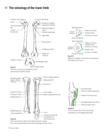
Fig.41.3
Diagram to explain the borders and surfaces
Patellar surface of the fibula (see text)
Medial epicondyle
Fig.41.1 Intercondylar fossa
The femur, anterior and posterior views
and the lower end from below
Intercondyle eminence
Medial condyle
Head of fibula Epiphyseal line
Tubercle
Soleal line
Peroneal nerve
Fibular collateral ligament
Lateral surface Vertical line
Anterior border Interosseous membrane
Tendon of biceps (cut)
Medial surface
Superficial peroneal nerve
Deep peroneal nerve
Fig.41.4
Medial malleolus The knee viewed from the lateral side
Talus showing the common peroneal nerve
Lateral malleolus
Sustentaculum tali
Attachment of
Fig.41.2 tendo calcaneus
The front and back of the tibia, fibula and ankle region.
The interosseous membrane and its openings are also shown
92 Lower limb