
Page 180 - Concise Pathology for Exam Preparation ( PDFDrive )
P. 180
7 Infections 165
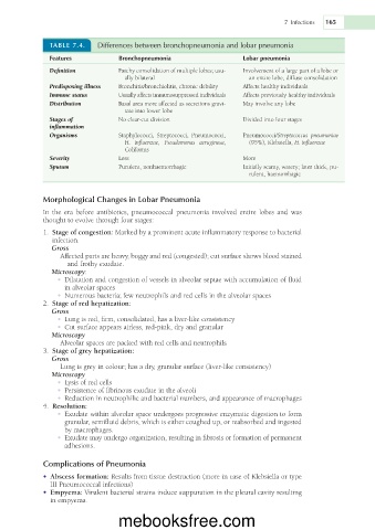
TABLE 7.4. Differences between bronchopneumonia and lobar pneumonia
Features Bronchopneumonia Lobar pneumonia
Definition Patchy consolidation of multiple lobes; usu- Involvement of a large part of a lobe or
ally bilateral an entire lobe, diffuse consolidation
Predisposing illness Bronchitis/bronchiolitis, chronic debility Affects healthy individuals
Immune status Usually affects immunosuppressed individuals Affects previously healthy individuals
Distribution Basal area more affected as secretions gravi- May involve any lobe
tate into lower lobe
Stages of No clear-cut division Divided into four stages
inflammation
Organisms Staphylococci, Streptococci, Pneumococci, Pneumococci/Streptococcus pneumoniae
H. influenzae, Pseudomonas aeruginosa, (95%), Klebsiella, H. influenzae
Coliforms
Severity Less More
Sputum Purulent, nonhaemorrhagic Initially scanty, watery; later thick, pu-
rulent, haemorrhagic
Morphological Changes in Lobar Pneumonia
In the era before antibiotics, pneumococcal pneumonia involved entire lobes and was
thought to evolve through four stages:
1. Stage of congestion: Marked by a prominent acute inflammatory response to bacterial
infection.
Gross
Affected parts are heavy, boggy and red (congested); cut surface shows blood stained
and frothy exudate.
Microscopy:
• Dilatation and congestion of vessels in alveolar septae with accumulation of fluid
in alveolar spaces
• Numerous bacteria; few neutrophils and red cells in the alveolar spaces
2. Stage of red hepatization:
Gross
• Lung is red, firm, consolidated, has a liver-like consistency
• Cut surface appears airless, red-pink, dry and granular
Microscopy
Alveolar spaces are packed with red cells and neutrophils
3. Stage of grey hepatization:
Gross
Lung is grey in colour; has a dry, granular surface (liver-like consistency)
Microscopy
• Lysis of red cells
• Persistence of fibrinous exudate in the alveoli
• Reduction in neutrophilic and bacterial numbers, and appearance of macrophages
4. Resolution:
• Exudate within alveolar space undergoes progressive enzymatic digestion to form
granular, semifluid debris, which is either coughed up, or reabsorbed and ingested
by macrophages.
• Exudate may undergo organization, resulting in fibrosis or formation of permanent
adhesions.
Complications of Pneumonia
• Abscess formation: Results from tissue destruction (more in case of Klebsiella or type
III Pneumococcal infections)
• Empyema: Virulent bacterial strains induce suppuration in the pleural cavity resulting
in empyema.
mebooksfree.com