
Page 188 - Concise Pathology for Exam Preparation ( PDFDrive )
P. 188
7 Infections 173
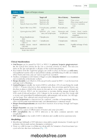
TABLE 7.5. Types of herpes viruses
Herpes
type Name Target cell Site of latency Transmission
1 HSV-1 Epithelial cells Neurons Close contact
2 HSV-2 Epithelial cells Neurons Close contact usually sexual
3 Varicella Zoster virus (VSV) Epithelial cells Neurons Contact or respiratory
route
4 Epstein–Barr virus (EBV) B lymphocytes, epithelial B lymphocytes Saliva
cells
5 Cytomegalovirus (CMV) Epithelial cells, mono- Monocytes and Contact, blood transfu-
cytes, and lymphocytes lymphocytes sions, transplantation,
congenital
6 Herpes lymphotropic T lymphocytes T lymphocytes Contact, respiratory route
virus
7 Human herpes virus-7 T lymphocytes T lymphocytes Unknown
(HHV-7)
8 Human herpes virus-8 Endothelial cells Unknown Possibly exchange of body
(HHV-8)/ fluids
Kaposi sarcoma-associated
herpes virus (KSHV)
Clinical Manifestations
• Oral herpes can be caused by HSV-1 or HSV-2. In primary herpetic gingivostomati-
tis, the typical clear lesions are the first to develop followed by ulcers. The infection
starts on the lips and spreads to all parts of the mouth and pharynx.
• Reactivation from the trigeminal ganglia can result in what are known as cold sores.
Intraepithelial vesicles (due to intracellular oedema and ballooning of cells) are formed,
which burst and crust, and can lead to superficial ulceration.
• Swollen, erythematous HSV lesions of fingers or palm (herpetic whitlow) occur in infants
and occasionally, in healthcare workers.
• Herpes keratitis is an infection of the eye primarily caused by HSV-1. It can be recurrent
and may lead to blindness.
• Genital herpes is usually the result of HSV-2 with about 10% of cases being the result
of HSV-1. Primary infection is often asymptomatic, but sometimes painful lesions can
develop on glans or shaft of the penis in men and on vulva, vagina, cervix and perianal
region of women. HSV-2 can be transmitted to neonates during passage through the
birth canal of infected mothers. HSV-2 disease in the neonate can vary from being mild
to severe with generalized lymphadenopathy, splenomegaly and necrotic foci through-
out the lungs, liver, adrenals and central nervous system.
• Eczema herpeticum is characterized by confluent, pustular or haemorrhagic blisters,
often with bacterial superinfection and viral dissemination to internal viscera.
• Herpes bronchopneumonia can result from insertion of an airway through oral herpes
lesions.
• Herpes hepatitis can cause liver failure.
• HSV can be a cause of inflammation of rectum and anus (proctitis).
• HSV encephalitis due to HSV-1 infection is the most common sporadic viral
encephalitis.
• HSV meningitis is the result of HSV-2 infection and usually resolves spontaneously.
Morphology
• Morphologic hallmark of HSV infection is large pink-to-purple intranuclear (Cowdry type A)
inclusions, which push nuclear chromatin to periphery.
• There is a mild increase in cellular size along with the formation of multinucleated syn-
cytial cells that also have inclusions (Fig. 7.8).
mebooksfree.com