
Page 192 - Concise Pathology for Exam Preparation ( PDFDrive )
P. 192
7 Infections 177
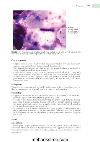
Budding
candidal yeast
forms forming a
pseudohyphae
FIGURE 7.9. Yeast forms of Candida (small, thin-walled ovoid cells of 4–6 microns that
reproduce by budding and form pseudohyphae; PAS stain; 4003).
Cryptococcosis
• Cryptococcosis is a rare fungal infection caused by inhalation of Cryptococcus neofor-
mans, an encapsulated fungus that is ordinarily found in soil.
• Once inhaled, the infection may heal on its own, remain localized in the lungs, or
spread throughout the body (dissemination).
• Cryptococcosis mostly occurs in immunocompromised individuals. In people with
normal immune system, the infection may have no symptoms. However, in people with
impaired immune systems, Cryptococcus may even spread to the brain (causing menin-
goencephalitis). Disseminated cryptococcosis usually involves the skin, liver, spleen,
adrenals and bones.
Pathogenesis
Virulence is due to capsular polysaccharides and enzymes, which prevent phagocytosis by
alveolar macrophages and inhibits leukocyte recruitment and migration.
Morphology
• Cryptococcus has yeast but no hyphal forms. It is 5–10 microns in size and has a thick
gelatinous capsule that is valuable for diagnosis (Fig. 7.10).
• Capsular polysaccharide stains intense red with periodic acid-Schiff (PAS) and mucicar-
mine stains in tissues, and can be detected with antibody-coated beads in an agglutina-
tion assay. India ink preparation gives a negative image, visualizing the thick capsule as
a clear halo, but not staining the yeast form.
• In immunosuppressed patients, organisms may evoke virtually no inflammatory reac-
tion, so gelatinous masses of fungi are seen in the tissue (gelatinous reaction). In nonim-
munosuppressed patients, the fungi induce a chronic granulomatous reaction. Suppura-
tion is rare.
Molds
Aspergillosis
This saprophytic fungus sporulates and produces conidia (asexual spores) that are readily
aerosolized. Molecular studies of Aspergillus isolated from opportunistic infections show
many different strains of Aspergillus, Aspergillus fumigatus is the most common species to
cause disease.
mebooksfree.com