
Page 193 - Concise Pathology for Exam Preparation ( PDFDrive )
P. 193
178 SECTION I General Pathology
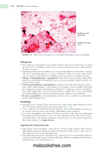
Budding in ovoid
yeast cells
Cryptococcal yeast
forms
FIGURE 7.10. Yeast forms of Cryptococcus neoformans showing a lot of size variation.
Pathogenesis
• The small size of Aspergillus spores enables them to reach alveoli where they are taken
up by alveolar macrophages, which secrete cytokines and chemokines to elicit adaptive
immune responses.
• Aspergillus produces several virulence factors, including adhesins, antioxidants, enzymes
and toxins. Aspergillus species is a source of aflatoxin, which is a major cause of liver
cancer in Africa. Sensitization to Aspergillus spores can produce an allergic alveolitis.
• Allergic bronchopulmonary aspergillosis results from hypersensitivity arising from
superficial colonization of bronchial mucosa and may eventually result in chronic ob-
structive lung disease.
• Colonizing aspergillosis (aspergilloma) is defined as growth of the fungus in pulmo-
nary cavities with minimal or no invasion of the tissues. Cavities usually result from
pre-existing tuberculosis, bronchiectasis, old infarcts or abscesses. Masses of fungal hy-
phae called fungus balls are seen lying free within the cavities. They may be surrounded
by minimal inflammatory reaction to marked chronic inflammation and fibrosis.
• Invasive aspergillosis is an opportunistic infection that is confined to immunosup-
pressed and debilitated hosts.
Morphology
• Aspergillus forms fruiting bodies (particularly in cavities) and septate filaments, which
are 5–10 microns thick and branch at acute angles (Fig. 7.11).
• It has a tendency to invade blood vessels; therefore, areas of haemorrhage and infarction
are usually superimposed on necrotizing, inflammatory tissue reactions.
• In invasive aspergillosis, the primary lesions are usually in the lung, but widespread
haematogenous dissemination is common. The pulmonary lesions take form of necro-
tizing pneumonia with sharply delineated, rounded, grey foci with haemorrhagic bor-
ders, often referred to as target lesions.
Zygomycosis (Mucormycosis)
• Zygomycetes form nonseptate, broad (6–50 microns) fungal hyphae with frequent
right-angled branching, which are readily demonstrated in the necrotic tissues by hae-
matoxylin and eosin or special fungal stains.
• Also called mucormycosis or phycomycosis, zygomycosis is an opportunistic infection
caused by ‘bread mold fungi’, including Rhizopus, Absidia, Cumunghanrella and Mucor,
which belong to the class Zygomycetes.
mebooksfree.com