
Page 196 - Concise Pathology for Exam Preparation ( PDFDrive )
P. 196
7 Infections 181
Eumycetoma
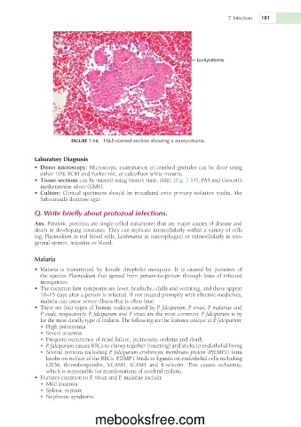
FIGURE 7.14. H&E-stained section showing a eumycetoma.
Laboratory Diagnosis
• Direct microscopy: Microscopic examination of crushed granules can be done using
either 10% KOH and Parker ink, or calcofluor white mounts.
• Tissue sections can be stained using Gram’s stain, H&E (Fig. 7.14), PAS and Grocott’s
methenamine silver (GMS).
• Culture: Clinical specimens should be inoculated onto primary isolation media, like
Sabouraud’s dextrose agar.
Q. Write briefly about protozoal infections.
Ans. Parasitic protozoa are single-celled eukaryotes that are major causes of disease and
death in developing countries. They can replicate intracellularly within a variety of cells
(eg, Plasmodium in red blood cells, Leishmania in macrophages) or extracellularly in uro-
genital system, intestine or blood.
Malaria
• Malaria is transmitted by female Anopheles mosquito. It is caused by parasites of
the species Plasmodium that spread from person-to-person through bites of infected
mosquitoes.
• The common first symptoms are fever, headache, chills and vomiting, and these appear
10–15 days after a person is infected. If not treated promptly with effective medicines,
malaria can cause severe illness that is often fatal.
• There are four types of human malaria caused by P. falciparum, P. vivax, P. malariae and
P. ovale, respectively. P. falciparum and P. vivax are the most common. P. falciparum is by
far the most deadly type of malaria. The following are the features unique to P. falciparum:
• High parasitemia
• Severe anaemia
• Frequent occurrence of renal failure, pulmonary oedema and death
• P. falciparum causes RBCs to clump together (rosetting) and sticks to endothelial lining
• Several proteins including P. falciparum erythrocyte membrane protein (PfEMP1) form
knobs on surface of the RBCs. PfEMP1 binds to ligands on endothelial cells including
CD36, thrombospondin, VCAM1, ICAM1 and E-selectin. This causes ischaemia,
which is responsible for manifestations of cerebral malaria.
• Features common to P. vivax and P. malariae include
• Mild anaemia
• Splenic rupture
• Nephrotic syndrome
mebooksfree.com