
Page 636 - Concise Pathology for Exam Preparation ( PDFDrive )
P. 636
23 The Central Nervous System 621
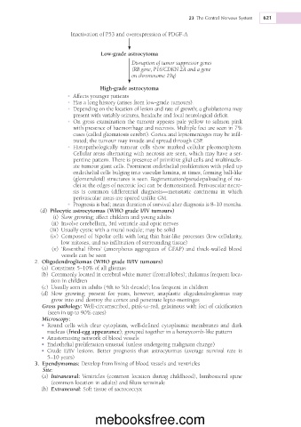
Inactivation of P53 and overexpression of PDGFA
Lowgrade astrocytoma
Disruption of tumor suppressor genes
(RB gene, P16/CDKN 2A and a gene
on chromosome 19q)
Highgrade astrocytoma
• Affects younger patients
• Has a long history (arises from low-grade tumours).
�
• Depending on the location of lesion and rate of growth; a glioblastoma may
�
present with variably seizures, headache and focal neurological deficit.
• On gross examination the tumour appears pale yellow to salmon pink
�
with presence of haemorrhage and necrosis. Multiple foci are seen in 7%
cases (called gliomatosis cerebri). Cortex and leptomeninges may be infil-
trated; the tumour may invade and spread through CSF.
• Histopathologically tumour cells show marked cellular pleomorphism.
�
Cellular areas alternating with necrosis are seen, which may have a ser-
pentine pattern. There is presence of primitive glial cells and multinucle-
ate tumour giant cells. Prominent endothelial proliferation with piled up
endothelial cells bulging into vascular lumina, at times, forming ball-like
(glomeruloid) structures is seen. Regimentation/pseudopalisading of nu-
clei at the edges of necrotic foci can be demonstrated. Perivascular necro-
sis is common (differential diagnosis—metastatic carcinoma in which
perivascular areas are spared unlike GM.
• Prognosis is bad; mean duration of survival after diagnosis is 8–10 months.
(d) �Pilocytic astrocytomas (WHO grade I/IV tumours)
(i) Slow growing; affect children and young adults
(ii) Involve cerebellum, 3rd ventricle and optic nerves
(iii) Usually cystic with a mural nodule; may be solid
(iv) Composed of bipolar cells with long thin hair-like processes (low cellularity,
low mitoses, and no infiltration of surrounding tissue)
(v) �‘Rosenthal fibres’ (amorphous aggregates of GFAP) and thick-walled blood
vessels can be seen
2. (WHO grade II/IV tumours)
Oligodendrogliomas
(a) Constitute 5–10% of all gliomas
(b) Commonly located in cerebral white matter (frontal lobes); thalamus frequent loca-
tion in children
(c) Usually seen in adults (4th to 5th decade); less frequent in children
(d) Slow growing; present for years, however, anaplastic oligodendrogliomas may
grow into and destroy the cortex and penetrate lepto-meninges
Gross pathology: Well-circumscribed, pink-to-red, gelatinous with foci of calcification
up to 90% cases)
(seen in
Microscopy:
• Round cells with clear cytoplasm, well-defined cytoplasmic membranes and dark
nucleus (fried-egg appearance); grouped together in a honeycomb-like pattern
• Anastomosing network of blood vessels
• Endothelial proliferation unusual (unless undergoing malignant change)
• Grade II/IV lesions. Better prognosis than astrocytomas (average survival rate is
5–10 years)
3. Ependymomas: Develop from lining of blood vessels and ventricles
Site:
Intraneural: Ventricles (common location during childhood), lumbosacral spine
(a) �
(common location in adults) and filum terminale
(b) �Extraneural: Soft tissue of sacrococcyx
mebooksfree.com