
Page 523 - Textbook of Pathology, 6th Edition
P. 523
507
Chapter 18 The Eye, ENT and Neck
Chapter 18
EYE
NORMAL STRUCTURE
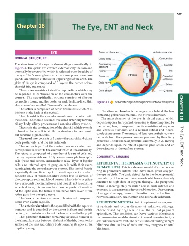
The structure of the eye is shown diagrammatically in
Fig. 18.1. The eyelids are covered externally by the skin and
internally by conjunctiva which is reflected over the globe of
the eye. The lacrimal glands which are compound racemose
glands are situated at the outer upper angle of the orbit. The
globe of the eye is composed of 3 layers: the cornea-sclera,
choroid-iris, and retina.
The cornea consists of stratified epithelium which may
be regarded as continuation of the conjunctiva over the
cornea. The subepithelial stroma consists of fibrous
connective tissue, and the posterior endothelium-lined thin Figure 18.1 Schematic diagram of longitudinal section of the eyeball.
elastic membrane called Descemet’s membrane.
The sclera is composed of dense fibrous tissue which is CHAPTER 18
thickest at the back of the eyeball. The vitreous chamber is the large space behind the lens
The choroid is the vascular membrane in contact with containing gelatinous material, the vitreous humour.
the sclera. The choroid becomes thickened anteriorly forming The main function of the eye is visual acuity which
ciliary body, ciliary processes and contains ciliary muscle. depends upon a transparent focussing system comprised by
The iris is the continuation of the choroid which extends the cornea, lens, transparent media consisting of aqueous
in front of the lens. It is similar in structure to the choroid and vitreous humours, and a normal retinal and neural
but contains pigment cells. conduction system. The cornea and lens receive their nutrient
The uveal tract consists of 3 parts—the choroid and ciliary demands from the aqueous humour produced by the ciliary
body posteriorly, and the iris anteriorly. processes. The intraocular pressure is normally 15-20 mmHg
The retina is part of the central nervous system and and depends upon the rate of aqueous production and on The Eye, ENT and Neck
corresponds in extent to the choroid which it lines internally. the resistance in the outflow system.
The retina is composed of a number of layers of cells and
their synapses which are of 3 types—external photoreceptor CONGENITAL LESIONS
cells (rods and cones), intermediate relay layer of bipolar
cells, and internal layer of ganglion cells with their axons RETROLENTAL FIBROPLASIA (RETINOPATHY OF
running into the central nervous system. The central fovea is PREMATURITY). This is a developmental disorder occur-
a specially differentiated spot in the retina posteriorly which ring in premature infants who have been given oxygen-
consists only of photosensitive cones but is devoid of therapy at birth. The basic defect lies in the developmental
photoreceptor rods and blood vessels. Macula lutea or yellow prematurity of the retinal blood vessels which are extremely
spot surrounds the central fovea and though not as sensitive sensitive to high dose of oxygen-therapy. The peripheral
as central fovea, it is more so than the other parts of the retina. retina is incompletely vascularised in such infants and
At the optic disc, the fibres of the nerve fibre layer of the exposure to oxygen results in vaso-obliteration. On stoppage
retina pass into the optic nerve. of oxygen-therapy, vasoproliferation begins leading to
The lens is the biconvex mass of laminated transparent neovascularisation, cicatrisation and retinal detachment.
tissue with elastic capsule. RETINITIS PIGMENTOSA. Retinitis pigmentosa is a group
The anterior chamber is the space filled with the aqueous of systemic and ocular diseases of unknown etiology,
humour, and is bounded by the cornea in front and the iris characterised by degeneration of the retinal pigment
behind, with anterior surface of the lens exposed in the pupil. epithelium. The condition can have various inheritance
The posterior chamber containing aqueous humour is patterns—autosomal dominant, autosomal recessive trait, or
the triangular space between the back of the iris, the anterior sex-linked recessive trait. The earliest clinical finding is night
surface of the lens and ciliary body forming its apex at the blindness due to loss of rods and may progress to total
pupillary margin. blindness.