
Page 524 - Textbook of Pathology, 6th Edition
P. 524
508
Histologically, there is disappearance of rods and cones
of the photoreceptor layer of the retina, degeneration of
retinal pigment epithelium and ingrowth of glial
membrane on the optic disc.
INFLAMMATORY CONDITIONS
Inflammatory conditions of the eye are designated according
to the tissue affected. ‘Uveitis’ is the commonly used term
for the ocular inflammation of the uveal tract which is the
most vascular tissue of the eye. However, specific designation
is used for the type of tissue of eye inflamed. Some of the
important types are described below.
STYE (HORDEOLUM). Stye or ‘external hordeolum’ is an
acute suppurative inflammation of the sebaceous glands of
Zeis, the apocrine glands of Moll and the eyelash follicles.
The less common ‘internal hordeolum’ is an acute
suppurative inflammation of the meibomian glands.
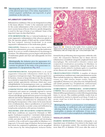
CHALAZION. Chalazion is a very common lesion and is Figure 18.2 Chalazion of the eyelid. There is presence of a
the chronic inflammatory process involving the meibomian lipogranuloma having central pool of fat and surrounded by mixed
glands. It occurs as a result of obstruction to the drainage of inflammatory cells and foreign body type multinucleated giant cells.
secretions. The inflammatory process begins with destruction
of meibomian glands and duct and subsequently involves formed by the proliferating microorganisms within the cells.
tarsal plate. Later, the conjunctiva thickens due to dense chronic
inflammatory cell infiltrate alongwith lymphoid follicles and
Histologically, the chalazion gives the appearance of a macrophages. The end-result is extensive corneal and
chronic inflammatory granuloma located in the tarsus and conjunctival cicatrisation accounting for blindness in
contains fat globules in the centre of the granulomas i.e. trachoma. Inclusion conjunctivitis, though caused by an
SECTION III
appearance of a lipogranuloma (Fig. 18.2). organism closely related to trachoma agent, is a much less
severe disease and causes mild keratoconjunctivitis.
ENDOPHTHALMITIS. Endophthalmitis is an acute
suppurative intraocular inflammation which may be of GRANULOMATOUS UVEITIS. A number of chronic
exogenous or endogenous origin. The exogenous agents may granulomatous conditions may cause granulomatous uveitis.
be bacteria, viruses or fungi introduced into the eye during These include bacteria (e.g. tuberculosis, leprosy, syphilis),
an accidental or surgical perforating wound. The endogenous viruses (e.g. CMV disease, herpes zoster), fungi (e.g.
agents include opportunistic infections which may cause aspergillosis, blastomycosis, phycomycosis, histoplasmosis),
endophthalmitis via haematogenous route e.g. candidiasis, and certain parasites (e.g. toxoplasmosis, onchocerciasis).
toxoplasmosis, nocardiosis, aspergillosis and cryptococcosis. Granulomatous uveitis is common in sarcoidosis as well.
CONJUNCTIVITIS AND KERATOCONJUNCTIVITIS. SYMPATHETIC OPHTHALMIA (SYMPATHETIC
Systemic Pathology
Conjunctiva and cornea are constantly exposed to various UVEITIS). This is an uncommon condition in which there is
types of physical, chemical, microbial (bacteria, fungi, bilateral diffuse granulomatous uveitis following penetrating
viruses) and allergic agents and hence prone to develop acute, injury to one eye. The condition probably results from an
subacute and chronic inflammations. In the acute stage, there autosensitivity reaction to injured uveal tissue. It leads to a
is corneal oedema and infiltration by inflammatory cells, severe visual loss in both the eyes if not diagnosed and
affecting the transparency of the cornea. In the more chronic treated early.
form of inflammation, there is proliferation of small blood Histologically, there is granulomatous uveal inflamma-
vessels in the normally avascular cornea and infiltration by tion consisting of epithelioid cells and lymphocytes
lymphocytes and plasma cells (pannus formation).
affecting both the eyes. There is no necrosis and no
TRACHOMA AND INCLUSION CONJUNCTIVITIS. neutrophilic or plasma cell infiltration. If lens is also
Both these conditions are caused by Chlamydia or TRIC injured, it results in phacoanaphylactic endophthalmitis.
agents. Trachoma is caused by C. trachomatis while inclusion
conjunctivitis is caused by C. oculogenitalis. Trachoma is VASCULAR LESIONS
widely prevalent in the underdeveloped and developing
countries of the world and is responsible for blindness on a DIABETIC RETINOPATHY. Diabetic retinopathy is an
large scale. In the early stage of infection, the trachoma agent important cause of blindness. It is related to the degree and
that infects the conjunctival epithelium, can be recognised duration of glycaemic control. The condition develops in
in the smears by the intracytoplasmic inclusion bodies more than 60% of diabetics 15-20 years after the onset of