
Page 525 - Textbook of Pathology, 6th Edition
P. 525
509
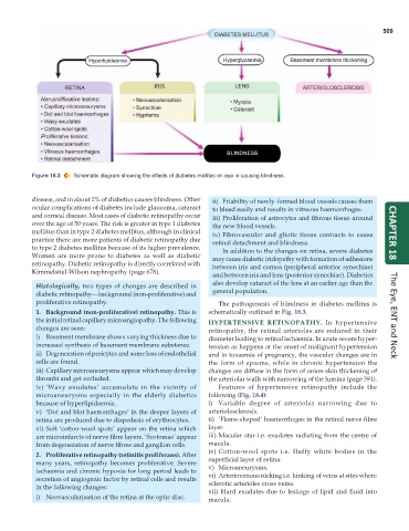
Figure 18.3 Schematic diagram showing the effects of diabetes mellitus on eye in causing blindness.
disease, and in about 2% of diabetics causes blindness. Other ii) Friability of newly-formed blood vessels causes them
ocular complications of diabetes include glaucoma, cataract to bleed easily and results in vitreous haemorrhages.
and corneal disease. Most cases of diabetic retinopathy occur iii) Proliferation of astrocytes and fibrous tissue around
over the age of 50 years. The risk is greater in type 1 diabetes the new blood vessels.
mellitus than in type 2 diabetes mellitus, although in clinical iv) Fibrovascular and gliotic tissue contracts to cause CHAPTER 18
practice there are more patients of diabetic retinopathy due retinal detachment and blindness.
to type 2 diabetes mellitus because of its higher prevalence. In addition to the changes on retina, severe diabetes
Women are more prone to diabetes as well as diabetic may cause diabetic iridopathy with formation of adhesions
retinopathy. Diabetic retinopathy is directly correlated with between iris and cornea (peripheral anterior synechiae)
Kimmelstiel-Wilson nephropathy (page 678).
and between iris and lens (posterior synechiae). Diabetics
also develop cataract of the lens at an earlier age than the
Histologically, two types of changes are described in
diabetic retinopathy—background (non-proliferative) and general population.
proliferative retinopathy. The pathogenesis of blindness in diabetes mellitus is
1. Background (non-proliferative) retinopathy. This is schematically outlined in Fig. 18.3.
the initial retinal capillary microangiopathy. The following HYPERTENSIVE RETINOPATHY. In hypertensive The Eye, ENT and Neck
changes are seen: retinopathy, the retinal arterioles are reduced in their
i) Basement membrane shows varying thickness due to diameter leading to retinal ischaemia. In acute severe hyper-
increased synthesis of basement membrane substance. tension as happens at the onset of malignant hypertension
ii) Degeneration of pericytes and some loss of endothelial and in toxaemia of pregnancy, the vascular changes are in
cells are found. the form of spasms, while in chronic hypertension the
iii) Capillary microaneurysms appear which may develop changes are diffuse in the form of onion-skin thickening of
thrombi and get occluded. the arteriolar walls with narrowing of the lumina (page 391).
iv) ‘Waxy exudates’ accumulate in the vicinity of Features of hypertensive retinopathy include the
microaneurysms especially in the elderly diabetics following (Fig. 18.4):
because of hyperlipidaemia. i) Variable degree of arteriolar narrowing due to
v) ‘Dot and blot haemorrhages’ in the deeper layers of arteriolosclerosis.
retina are produced due to diapedesis of erythrocytes. ii) ‘Flame-shaped’ haemorrhages in the retinal nerve fibre
vi) Soft ‘cotton-wool spots’ appear on the retina which layer.
are microinfarcts of nerve fibre layers. ‘Scotomas’ appear iii) Macular star i.e. exudates radiating from the centre of
from degeneration of nerve fibres and ganglion cells. macula.
iv) Cotton-wool spots i.e. fluffy white bodies in the
2. Proliferative retinopathy (retinitis proliferans). After superficial layer of retina.
many years, retinopathy becomes proliferative. Severe v) Microaneurysms.
ischaemia and chronic hypoxia for long period leads to vi) Arteriovenous nicking i.e. kinking of veins at sites where
secretion of angiogenic factor by retinal cells and results sclerotic arterioles cross veins.
in the following changes: vii) Hard exudates due to leakage of lipid and fluid into
i) Neovascularisation of the retina at the optic disc. macula.