
Page 526 - Textbook of Pathology, 6th Edition
P. 526
510 choroid, exudation and haemorrhage under the retina
which may eventually get organised and heal by fibrosis
and result in permanent loss of central vision.
RETINAL DETACHMENT. Retinal detachment is the
separation of the neurosensory retina from the retinal
pigment epithelium. It may occur spontaneously in older
individuals past 50 years of age, or may be secondary to
trauma in the region of head and neck. Normally, the rods
and cones of the photoreceptor layer are interdigitated with
projections of the retinal pigment epithelium, but the two
can separate readily in some disease processes. There are 3
pathogenetic mechanisms of retinal detachment:
i) Pathologic processes in the vitreous or anterior segment,
causing traction on the retina.
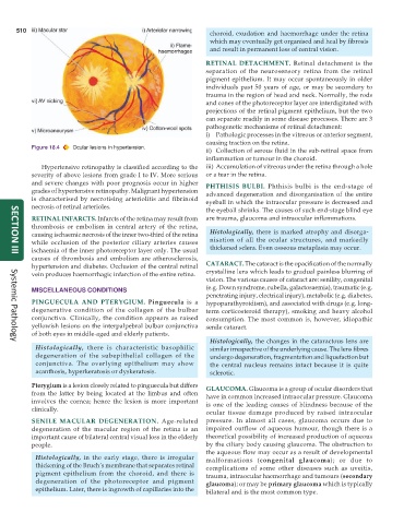
Figure 18.4 Ocular lesions in hypertension.
ii) Collection of serous fluid in the sub-retinal space from
inflammation or tumour in the choroid.
Hypertensive retinopathy is classified according to the iii) Accumulation of vitreous under the retina through a hole
severity of above lesions from grade I to IV. More serious or a tear in the retina.
and severe changes with poor prognosis occur in higher PHTHISIS BULBI. Phthisis bulbi is the end-stage of
grades of hypertensive retinopathy. Malignant hypertension advanced degeneration and disorganisation of the entire
is characterised by necrotising arteriolitis and fibrinoid eyeball in which the intraocular pressure is decreased and
necrosis of retinal arterioles.
the eyeball shrinks. The causes of such end-stage blind eye
RETINAL INFARCTS. Infarcts of the retina may result from are trauma, glaucoma and intraocular inflammations.
thrombosis or embolism in central artery of the retina,
causing ischaemic necrosis of the inner two-third of the retina Histologically, there is marked atrophy and disorga-
while occlusion of the posterior ciliary arteries causes nisation of all the ocular structures, and markedly
ischaemia of the inner photoreceptor layer only. The usual thickened sclera. Even osseous metaplasia may occur.
SECTION III
causes of thrombosis and embolism are atherosclerosis,
hypertension and diabetes. Occlusion of the central retinal CATARACT. The cataract is the opacification of the normally
vein produces haemorrhagic infarction of the entire retina. crystalline lens which leads to gradual painless blurring of
vision. The various causes of cataract are: senility, congenital
(e.g. Down syndrome, rubella, galactosaemia), traumatic (e.g.
MISCELLANEOUS CONDITIONS
penetrating injury, electrical injury), metabolic (e.g. diabetes,
PINGUECULA AND PTERYGIUM. Pinguecula is a hypoparathyroidism), and associated with drugs (e.g. long-
degenerative condition of the collagen of the bulbar term corticosteroid therapy), smoking and heavy alcohol
conjunctiva. Clinically, the condition appears as raised consumption. The most common is, however, idiopathic
yellowish lesions on the interpalpebral bulbar conjunctiva senile cataract.
of both eyes in middle-aged and elderly patients.
Histologically, the changes in the cataractous lens are
Systemic Pathology
Histologically, there is characteristic basophilic similar irrespective of the underlying cause. The lens fibres
degeneration of the subepithelial collagen of the undergo degeneration, fragmentation and liquefaction but
conjunctiva. The overlying epithelium may show the central nucleus remains intact because it is quite
acanthosis, hyperkeratosis or dyskeratosis. sclerotic.
Pterygium is a lesion closely related to pinguecula but differs GLAUCOMA. Glaucoma is a group of ocular disorders that
from the latter by being located at the limbus and often have in common increased intraocular pressure. Glaucoma
involves the cornea; hence the lesion is more important is one of the leading causes of blindness because of the
clinically.
ocular tissue damage produced by raised intraocular
SENILE MACULAR DEGENERATION. Age-related pressure. In almost all cases, glaucoma occurs due to
degeneration of the macular region of the retina is an impaired outflow of aqueous humour, though there is a
important cause of bilateral central visual loss in the elderly theoretical possibility of increased production of aqueous
people. by the ciliary body causing glaucoma. The obstruction to
the aqueous flow may occur as a result of developmental
Histologically, in the early stage, there is irregular malformations (congenital glaucoma); or due to
thickening of the Bruch’s membrane that separates retinal complications of some other diseases such as uveitis,
pigment epithelium from the choroid, and there is trauma, intraocular haemorrhage and tumours (secondary
degeneration of the photoreceptor and pigment glaucoma); or may be primary glaucoma which is typically
epithelium. Later, there is ingrowth of capillaries into the bilateral and is the most common type.