
Page 527 - Textbook of Pathology, 6th Edition
P. 527
There are 2 types of primary glaucoma—primary open- TABLE 18.1: Tumours and Tumour-like Lesions of the Eye 511
angle (chronic simple glaucoma) and primary angle-closure and Adnexal Structures.
(acute congestive glaucoma). Primary open-angle glaucoma
is more common type and is usually a genetically-determined Benign Malignant
disease. Primary angle-closure glaucoma occurs due to I. EYELID
shallow anterior chamber and hence narrow angle causing Squamous cell papilloma Squamous cell carcinoma
blockage to aqueous outflow. Basal cell papilloma Basal cell carcinoma
In all types of glaucoma, degenerative changes appear Sebaceous adenoma Sebaceous adenocarcinoma
after some duration and eventually damage to the optic nerve Naevi Malignant melanoma
and retina occurs. II. CONJUNCTIVA-CORNEA
Squamous cell papilloma Squamous cell carcinoma
PAPILLOEDEMA. Papilloedema is oedema of the optic disc Pseudoepitheliomatous Mucoepidermoid carcinoma
resulting from increased intracranial pressure. This is due hyperplasia
to anatomic continuation of the subarachnoid space of the Naevi Malignant melanoma
brain around the optic nerve so that raised intracranial Haemangioma
III. LACRIMAL GLAND
pressure is passed onto the optic disc area. In acute
papilloedema, there is oedema, congestion and haemorrhage Pleomorphic adenoma Carcinoma in pleomorphic
adenoma
at the optic disc. In chronic papilloedema, there is degene- IV. ORBIT
ration of nerve fibres, gliosis and optic atrophy.
Glioma Malignant glioma
SJÖGREN’S SYNDROME. Sjögren’s syndrome is charac- Inflammatory ‘pseudotumour’ Malignant lymphoma
terised by triad of keratoconjunctivitis sicca, xerostomia (sicca Meningioma Rhabdomyosarcoma
syndrome) and rheumatoid arthritis. The condition occurs V. INTRAOCULAR
due to immunologically-mediated destruction of the lacrimal Naevi Malignant melanoma
and salivary glands along- with another autoimmune disease Neurofibroma Retinoblastoma
(Chapter 4).
is seen more commonly in the upper eyelid (basal cell
MIKULICZ’S SYNDROME. This is characterised by
inflammatory enlargement of lacrimal and salivary glands carcinoma is seen more frequently in the lower eyelid). CHAPTER 18
(Chapter 19). The condition may occur with Sjögren’s MORPHOLOGIC FEATURES. Grossly, the tumour
syndrome, or with some diseases like sarcoidosis, leukaemia, appears as a localised or diffuse swelling of the tarsus, or
lymphoma and macroglobulinaemia. may be in the form of ulcerated or papillomatous tumour
at the lid margin.
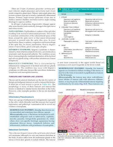
TUMOURS AND TUMOUR-LIKE LESIONS Microscopically, the tumour may show well-differen-
The eye and its adnexal structures are the site of a variety of tiated lobules of tumour cells with sebaceous differen-
benign and malignant tumours as well as tumour-like lesions. tiation, or may be poorly-differentiated tumour requiring
A brief list of such lesions is given in Table 18.1. The confirmation by fat stains (Fig. 18.5). These tumours can
morphology of many of these tumours and tumour-like
lesions is identical to similar lesions elsewhere in the body. The Eye, ENT and Neck
However, a few examples peculiar to the eye are described
below.
Inflammatory Pseudotumours
These are a group of inflammatory enlargements, especially
in the orbit, which clinically look like tumours but surgical
exploration and pathologic examination fail to reveal any
evidence of neoplasm.
MORPHOLOGIC FEATURES. Grossly, these lesions are
circumscribed and sometimes have fibrous capsule.
Microscopically, many of the lesions can be placed in well-
established categories such as tuberculous, syphilitic,
mycotic, parasitic, foreign-body granuloma etc, while
others show non-specific histologic appearance having
abundant fibrous tissue, lymphoid follicles and
inflammatory infiltrate with prominence of eosinophils.
Sebaceous Carcinoma
This is the most frequent tumour of the eyelid next only to basal
cell carcinoma, although it is very rare tumour elsewhere in Figure 18.5 Sebaceous carcinoma. The tumour cells are arranged
as lobules with peripheral basaloid cells and pale cells in the centre. The
the body. It arises either from the meibomian glands in the cells have sebaceous differentiation appreciated by foamy, vacuolated
tarsus or from Zeis’ glands of eyelash follicles. The tumour cytoplasm.