
Page 528 - Textbook of Pathology, 6th Edition
P. 528
512
Figure 18.6 Choroidal melanoma appearing as a pigmented mass
pushing the retina forward over it.
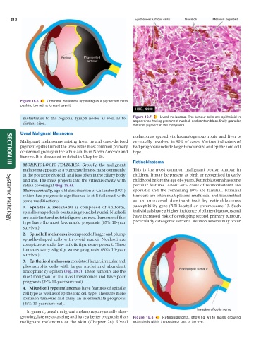
metastasise to the regional lymph nodes as well as to Figure 18.7 Uveal melanoma. The tumour cells are epithelioid in
distant sites. appearance having prominent nucleoli and contain black finely granular
melanin pigment in the cytoplasm.
Uveal Malignant Melanoma
melanomas spread via haematogenous route and liver is
Malignant melanomas arising from neural crest-derived eventually involved in 90% of cases. Various indicators of
pigment epithelium of the uvea is the most common primary bad prognosis include large tumour size and epithelioid cell
ocular malignancy in the white adults in North America and type.
Europe. It is discussed in detail in Chapter 26.
Retinoblastoma
MORPHOLOGIC FEATURES. Grossly, the malignant
SECTION III
melanoma appears as a pigmented mass, most commonly This is the most common malignant ocular tumour in
in the posterior choroid, and less often in the ciliary body children. It may be present at birth or recognised in early
and iris. The mass projects into the vitreous cavity with childhood before the age of 4 years. Retinoblastoma has some
retina covering it (Fig. 18.6). peculiar features. About 60% cases of retinoblastoma are
Microscopically, age-old classification of Callender (1931) sporadic and the remaining 40% are familial. Familial
which has prognostic significance is still followed with tumours are often multiple and multifocal and transmitted
some modifications: as an autosomal dominant trait by retinoblastoma
1. Spindle A melanoma is composed of uniform, susceptibility gene (RB) located on chromosome 13. Such
spindle-shaped cells containing spindled nuclei. Nucleoli individuals have a higher incidence of bilateral tumours and
are indistinct and mitotic figures are rare. Tumours of this have increased risk of developing second primary tumour,
type have the most favourable prognosis (85% 10-year particularly osteogenic sarcoma. Retinoblastoma may occur
Systemic Pathology
survival).
2. Spindle B melanoma is composed of larger and plump
spindle-shaped cells with ovoid nuclei. Nucleoli are
conspicuous and a few mitotic figures are present. These
tumours carry slightly worse prognosis (80% 10-year
survival).
3. Epithelioid melanoma consists of larger, irregular and
pleomorphic cells with larger nuclei and abundant
acidophilic cytoplasm (Fig. 18.7). These tumours are the
most malignant of the uveal melanomas and have poor
prognosis (35% 10-year survival).
4. Mixed cell type melanomas have features of spindle
cell type as well as of epithelioid cell type. These are more
common tumours and carry an intermediate prognosis
(45% 10-year survival).
In general, uveal malignant melanomas are usually slow-
growing, late metastasising and have a better prognosis than Figure 18.8 Retinoblastoma, showing white mass growing
malignant melanoma of the skin (Chapter 26). Uveal extensively within the posterior part of the eye.