
Page 529 - Textbook of Pathology, 6th Edition
P. 529
513
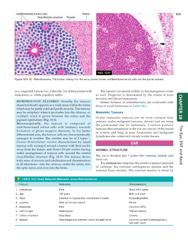
Figure 18.9 Retinoblastoma. The tumour arising from the retina (arrow) shows undifferentiated retinal cells and the typical rosettes.
as a congenital tumour too. Clinically, the child presents with The tumour can spread widely via haematogenous route
leukokoria i.e. white pupillary reflex. as well. Prognosis is determined by the extent of local
invasion and distant metastasis.
MORPHOLOGIC FEATURES. Grossly, the tumour Salient features of retinoblastoma are contrasted with
characteristically appears as a white mass within the retina those of uveal melanoma in Table 18.2.
which may be partly solid and partly necrotic. The tumour CHAPTER 18
may be endophytic when it protrudes into the vitreous, or Metastatic Tumours
exophytic when it grows between the retina and the Ocular metastatic tumours are far more common than
pigment epithelium (Fig. 18.8). primary ocular malignant tumours, choroid and iris being
Microscopically, the tumour is composed of the preferential site for metastasis. Common primary
undifferentiated retinal cells with tendency towards tumours that metastasise in the eye are cancers of the breast
formation of photo-receptor elements. In the better in women and lung in men. Leukaemia and malignant
differentiated area, the tumour cells are characteristically lymphoma also commonly invade ocular tissues.
arranged in rosettes. The rosettes may be of 2 types—
Flexner-Wintersteiner rosettes characterised by small EAR
tumour cells arranged around a lumen with their nuclei The Eye, ENT and Neck
away from the lumen, and Homer-Wright rosettes having NORMAL STRUCTURE
radial arrangement of tumour cells around the central
neurofibrillar structure (Fig. 18.9). The tumour shows The ear is divided into 3 parts—the external, middle and
wide areas of necrosis and calcification and dissemination inner ear.
in all directions—into the vitreous, under the retina, into The external ear comprises the auricle or pinna composed
the optic nerve and even into the brain. of cartilage, the external cartilaginous meatus and the
external bony meatus. The external meatus is lined by
TABLE 18.2: Uveal Malignant Melanoma versus Retinoblastoma.
Feature Melanoma Retinoblastoma
1. Inheritance Rare About 40% cases
2. Age >50 years Birth to 4 years
3. Race Common in Caucasians, uncommon in blacks No predisposition
4. Location Most commonly choroid Retina
5. Bilaterality Rare Common (30%)
6. Cell of origin Melanocytes Retinal neurons
7. Colour of tumour Grey-black Creamy
8. Spread Haematogenous common, rarely via optic nerve Common via both haematogenous
and optic nerve