
Page 532 - Textbook of Pathology, 6th Edition
P. 532
516 respiratory epithelium which may show squamous
metaplasia (Fig. 18.11). Nasal polyps may have
superimposed fungal infection.
RHINOSPORIDIOSIS. Rhinosporidiosis is caused by a
fungus, Rhinosporidium seeberi. Typically it occurs in a nasal
polyp but may be found in other locations like nasopharynx,
larynx and conjunctiva. The disease is common in India and
Sri Lanka and sporadic in other parts of the world.
Microscopically, besides the structure of inflammatory
or allergic polyp, large number of organisms of the size
of erythrocytes with chitinous wall are seen in the thick-
walled sporangia. Each sporangium may contain a few
thousand spores. On rupture of a sporangium, the spores
are discharged into the submucosa or on to the surface of
the mucosa. The intervening tissue consists of inflam-
matory granulation tissue (plasma cells, lymphocytes,
histiocytes, neutrophils) while the overlying epithelium
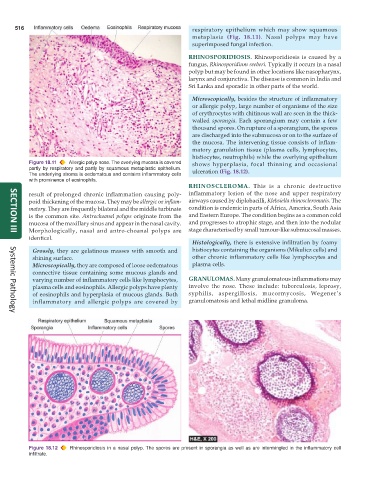
Figure 18.11 Allergic polyp nose. The overlying mucosa is covered shows hyperplasia, focal thinning and occasional
partly by respiratory and partly by squamous metaplastic epithelium. ulceration (Fig. 18.12).
The underlying stroma is oedematous and contains inflammatory cells
with prominence of eosinophils.
RHINOSCLEROMA. This is a chronic destructive
result of prolonged chronic inflammation causing poly- inflammatory lesion of the nose and upper respiratory
poid thickening of the mucosa. They may be allergic or inflam- airways caused by diplobacilli, Klebsiella rhinoscleromatis. The
matory. They are frequently bilateral and the middle turbinate condition is endemic in parts of Africa, America, South Asia
is the common site. Antrochoanal polyps originate from the and Eastern Europe. The condition begins as a common cold
mucosa of the maxillary sinus and appear in the nasal cavity. and progresses to atrophic stage, and then into the nodular
Morphologically, nasal and antro-choanal polyps are stage characterised by small tumour-like submucosal masses.
SECTION III
identical.
Histologically, there is extensive infiltration by foamy
Grossly, they are gelatinous masses with smooth and histiocytes containing the organisms (Mikulicz cells) and
shining surface. other chronic inflammatory cells like lymphocytes and
Microscopically, they are composed of loose oedematous plasma cells.
connective tissue containing some mucous glands and
varying number of inflammatory cells like lymphocytes, GRANULOMAS. Many granulomatous inflammations may
plasma cells and eosinophils. Allergic polyps have plenty involve the nose. These include: tuberculosis, leprosy,
of eosinophils and hyperplasia of mucous glands. Both syphilis, aspergillosis, mucormycosis, Wegener’s
inflammatory and allergic polyps are covered by granulomatosis and lethal midline granuloma.
Systemic Pathology
Figure 18.12 Rhinosporidiosis in a nasal polyp. The spores are present in sporangia as well as are intermingled in the inflammatory cell
infiltrate.