
Page 534 - Textbook of Pathology, 6th Edition
P. 534
518 crypts are filled with debris and pus giving it follicular
appearance. Chronic tonsillitis is caused by repeated attacks
of acute tonsillitis in which case the tonsils are small and
fibrosed. Acute tonsillitis may pass on to tissues adjacent to
tonsils to form peritonsillar abscess or quinsy.
PERITONSILLAR ABSCESS (QUINSY). Peritonsillar
abscess or quinsy occurs as a complication of acute tonsil-
litis. The causative organisms are staphylococci or
streptococci which are associated with infection of the tonsils.
The patient complains of acute pain in the throat, trismus,
difficulty in speech and inability to swallow. The glands
behind the angle of the mandible are enlarged and tender.
Besides the surgical management of the abscess, the patient
must be advised tonsillectomy because quinsy is frequently
recurrent.
RETROPHARYNGEAL ABSCESS. Formation of abscess in
the soft tissue between the posterior wall of the pharynx and
the vertebral column is called retropharyngeal abscess. It
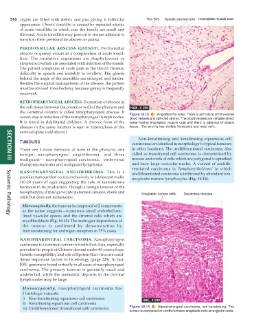
occurs due to infection of the retropharyngeal lymph nodes. Figure 18.13 Angiofibroma nose. There is admixture of thin-walled
blood vessels and spindled stroma. The blood vessels are variable-sized,
It is found in debilitated children. A chronic form of the some having incomplete muscle coat and there is absence of elastic
abscess in the same location is seen in tuberculosis of the tissue. The stroma has stellate fibroblasts and mast cells.
cervical spine (cold abscess).
Non-keratinising and keratinising squamous cell
TUMOURS carcinomas are identical in morphology to typical tumours
There are 4 main tumours of note in the pharynx, one in other locations. The undifferentiated carcinoma, also
benign—nasopharyngeal angiofibroma, and three called as transitional cell carcinoma, is characterised by
malignant— nasopharyngeal carcinoma, embryonal masses and cords of cells which are polygonal to spindled
SECTION III
rhabdomyosarcoma and malignant lymphoma. and have large vesicular nuclei. A variant of undiffe-
rentiated carcinoma is ‘lymphoepithelioma’ in which
NASOPHARYNGEAL ANGIOFIBROMA. This is a undifferentiated carcinoma is infiltrated by abundant non-
peculiar tumour that occurs exclusively in adolescent males neoplastic mature lymphocytes (Fig. 18.14).
(10-20 years of age) suggesting the role of testosterone
hormone in its production. Though a benign tumour of the
nasopharynx, it may grow into paranasal sinuses, cheek and
orbit but does not metastasise.
Microscopically, the tumour is composed of 2 components
as the name suggests—numerous small endothelium-
lined vascular spaces and the stromal cells which are
Systemic Pathology
myofibroblasts (Fig. 18.13). The androgen-dependence of
the tumour is confirmed by demonstration by
immunostaining for androgen receptors in 75% cases.
NASOPHARYNGEAL CARCINOMA. Nasopharyngeal
carcinoma is a common cancer in South-East Asia, especially
prevalent in people of Chinese descent under 45 years of age.
Genetic susceptibility and role of Epstein-Barr virus are consi-
dered important factors in its etiology (page 225). In fact,
EBV-genome is found virtually in all cases of nasopharyngeal
carcinoma. The primary tumour is generally small and
undetected, while the metastatic deposits in the cervical
lymph nodes may be large.
Microscopically, nasopharyngeal carcinoma has
3 histologic variants:
i) Non-keratinising squamous cell carcinoma
ii) Keratinising squamous cell carcinoma
iii) Undifferentiated (transitional cell) carcinoma Figure 18.14 Nasopharyngeal carcinoma, non-keratinising. The
tumour is composed of undifferentiated anaplastic cells arranged in nests.