
Page 539 - Textbook of Pathology, 6th Edition
P. 539
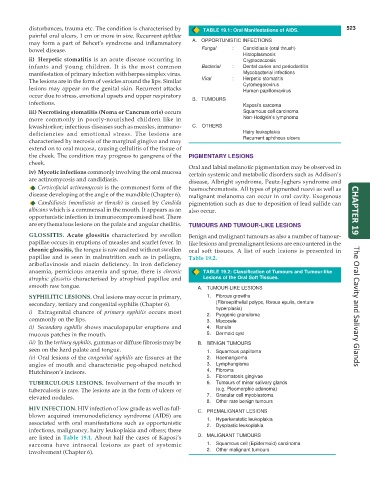
disturbances, trauma etc. The condition is characterised by TABLE 19.1: Oral Manifestations of AIDS. 523
painful oral ulcers, 1 cm or more in size. Recurrent aphthae
may form a part of Behcet’s syndrome and inflammatory A. OPPORTUNISTIC INFECTIONS
bowel disease. Fungal : Candidiasis (oral thrush)
Histoplasmosis
ii) Herpetic stomatitis is an acute disease occurring in Cryptococcosis
infants and young children. It is the most common Bacterial : Dental caries and periodontitis
manifestation of primary infection with herpes simplex virus. Mycobacterial infections
The lesions are in the form of vesicles around the lips. Similar Viral : Herpetic stomatitis
lesions may appear on the genital skin. Recurrent attacks Cytomegalovirus
Human papillomavirus
occur due to stress, emotional upsets and upper respiratory
infections. B. TUMOURS Kaposi’s sarcoma
iii) Necrotising stomatitis (Noma or Cancrum oris) occurs Squamous cell carcinoma
more commonly in poorly-nourished children like in Non-Hodgkin’s lymphoma
kwashiorkor; infectious diseases such as measles, immuno- C. OTHERS
deficiencies and emotional stress. The lesions are Hairy leukoplakia
characterised by necrosis of the marginal gingiva and may Recurrent aphthous ulcers
extend on to oral mucosa, causing cellulitis of the tissue of
the cheek. The condition may progress to gangrene of the PIGMENTARY LESIONS
cheek.
Oral and labial melanotic pigmentation may be observed in
iv) Mycotic infections commonly involving the oral mucosa certain systemic and metabolic disorders such as Addison’s
are actinomycosis and candidiasis. disease, Albright syndrome, Peutz-Jeghers syndrome and
Cervicofacial actinomycosis is the commonest form of the haemochromatosis. All types of pigmented naevi as well as
disease developing at the angle of the mandible (Chapter 6). malignant melanoma can occur in oral cavity. Exogenous
Candidiasis (moniliasis or thrush) is caused by Candida pigmentation such as due to deposition of lead sulfide can
albicans which is a commensal in the mouth. It appears as an also occur. CHAPTER 19
opportunistic infection in immunocompromised host. There
are erythematous lesions on the palate and angular cheilitis. TUMOURS AND TUMOUR-LIKE LESIONS
GLOSSITIS. Acute glossitis characterised by swollen Benign and malignant tumours as also a number of tumour-
papillae occurs in eruptions of measles and scarlet fever. In like lesions and premalignant lesions are encountered in the
chronic glossitis, the tongue is raw and red without swollen oral soft tissues. A list of such lesions is presented in
papillae and is seen in malnutrition such as in pellagra, Table 19.2.
ariboflavinosis and niacin deficiency. In iron deficiency
anaemia, pernicious anaemia and sprue, there is chronic TABLE 19.2: Classification of Tumours and Tumour-like
atrophic glossitis characterised by atrophied papillae and Lesions of the Oral Soft Tissues.
smooth raw tongue. A. TUMOUR-LIKE LESIONS
SYPHILITIC LESIONS. Oral lesions may occur in primary, 1. Fibrous growths
secondary, tertiary and congenital syphilis (Chapter 6). (Fibroepithelial polyps, fibrous epulis, denture The Oral Cavity and Salivary Glands
i) Extragenital chancre of primary syphilis occurs most 2. hyperplasia)
Pyogenic granuloma
commonly on the lips. 3. Mucocele
ii) Secondary syphilis shows maculopapular eruptions and 4. Ranula
mucous patches in the mouth. 5. Dermoid cyst
iii) In the tertiary syphilis, gummas or diffuse fibrosis may be B. BENIGN TUMOURS
seen on the hard palate and tongue. 1. Squamous papilloma
iv) Oral lesions of the congenital syphilis are fissures at the 2. Haemangioma
angles of mouth and characteristic peg-shaped notched 3. Lymphangioma
Hutchinson’s incisors. 4. Fibroma
5. Fibromatosis gingivae
TUBERCULOUS LESIONS. Involvement of the mouth in 6. Tumours of minor salivary glands
tuberculosis is rare. The lesions are in the form of ulcers or (e.g. Pleomorphic adenoma)
elevated nodules. 7. Granular cell myoblastoma
8. Other rare benign tumours
HIV INFECTION. HIV infection of low grade as well as full- C. PREMALIGNANT LESIONS
blown acquired immunodeficiency syndrome (AIDS) are
associated with oral manifestations such as opportunistic 1. Hyperkeratotic leukoplakia
2.
Dysplastic leukoplakia
infections, malignancy, hairy leukoplakia and others; these
are listed in Table 19.1. About half the cases of Kaposi’s D. MALIGNANT TUMOURS
sarcoma have intraoral lesions as part of systemic 1. Squamous cell (Epidermoid) carcinoma
involvement (Chapter 6). 2. Other malignant tumours