
Page 543 - Textbook of Pathology, 6th Edition
P. 543
527
Figure 19.5 Squamous cell (Epidermoid) carcinoma of oral cavity, patterns of gross appearance.
i) Ulcerative type—is the most frequent type and is of very well-differentiated squamous epithelium with
characterised by indurated ulcer and firm everted or rolled minimal atypia and hence has very good prognosis.
edges.
ii) Papillary or verrucous type—is soft and wart-like OTHER MALIGNANT TUMOURS
growth. Other less common malignant neoplasms which may be
iii) Nodular type—appears as a firm, slow growing encountered in the oral cavity are: malignant melanoma,
submucosal nodule. lymphoepithelial carcinoma, malignant lymphoma,
iv) Scirrhous type—is characterised by infiltration into malignant tumours of minor salivary glands, and various
deeper structures. sarcomas like rhabdomyosarcoma, liposarcoma, alveolar soft
All these types may appear on a background of part sarcoma, Kaposi’s sarcoma and fibrosarcoma. Metastatic
leukoplakia or erythroplasia of the oral mucosa. Enlarged tumours can also occur in the soft tissues of the mouth.
cervical lymph nodes may sometimes be present.
Histologically, squamous cell carcinoma ranges from TEETH AND PERIODONTAL TISSUES
well-differentiated keratinising carcinoma to highly- Although care of the teeth belongs to the field of dental CHAPTER 19
undifferentiated neoplasm (Chapter 26). Changes of profession, the fully educated medical doctor should be
epithelial dysplasia are often present in the surrounding familiar with certain principal diseases of teeth and
areas of the lesion. Carcinoma of the lip and intraoral periodontal tissues, especially about dental caries, periapical
squamous carcinoma are usually always well- abscess and periodontitis, and common cysts and
differentiated (Fig. 19.6).
odontogenic tumours of the jaw. But first, a brief account of
normal structure of these tissues is presented.
Carcinoma of the lip has a more favourable prognosis
due to visible and easily accessible location and less frequent NORMAL STRUCTURE
metastasis to the regional lymph nodes. However, intraoral
squamous carcinomas have poor prognosis because they are The teeth are normally composed of 3 calcified tissues,
detected late and metastasis to regional lymph nodes occur namely: enamel, dentin and cementum; and the pulp which is
early, especially in the case of carcinoma of tongue and soft composed of connective tissue. The teeth are peculiar than
palate. Verrucous carcinoma, on the other hand, is composed other calcified tissues of the body by being surrounded by The Oral Cavity and Salivary Glands
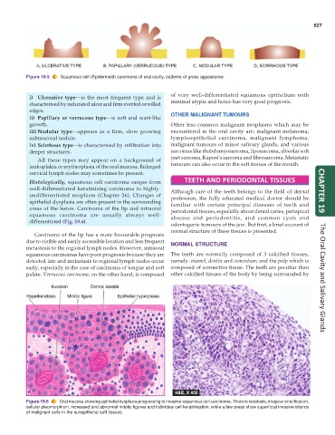
Figure 19.6 Oral mucosa showing epithelial dysplasia progressing to invasive squamous cell carcinoma. There is keratosis, irregular stratification,
cellular pleomorphism, increased and abnormal mitotic figures and individual cell keratinisation, while a few areas show superficial invasive islands
of malignant cells in the subepithelial soft tissues.