
Page 546 - Textbook of Pathology, 6th Edition
P. 546
530
Figure 19.9 Dental (Radicular) cyst. The cyst wall is composed of fibrous
tissue and is lined by non-keratinised squamous epithelium. The cyst wall is
densely infiltrated by chronic inflammatory cells, chiefly lymphocytes, plasma
cells and macrophages.
B. DEVELOPMENTAL CYSTS Non-odontogenic and Fissural Cysts
Odontogenic Cysts NASOPALATINE DUCT (INCISIVE CANAL, MEDIAN,
DENTIGEROUS (FOLLICULAR) CYST. Dentigerous cyst ANTERIOR MAXILLARY) CYST. This is the most common
arises from enamel of an unerupted tooth. The mandibular non-odontogenic (fissural) cyst and arises from the epithelial
SECTION III
third molars and the maxillary canines are most often remnants of the nasopalatine duct.
involved. Dentigerous cysts are less common than radicular Histologically, the cyst is lined by stratified squamous
cysts and occur more commonly in children and young epithelium, respiratory epithelium, or both.
individuals. These cysts are more significant because of
reported occurrence of ameloblastoma and carcinoma in NASOLABIAL (NASOALVEOLAR) CYST. This cyst is
them. situated in the soft tissues at the junction of median nasal,
lateral nasal and maxillary processes, at the ala of the nose,
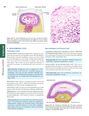
Histologically, dentigerous cyst is composed of a thin and sometimes extending into the nostril.
fibrous tissue wall lined by stratified squamous
epithelium. Thus, the cyst may resemble radicular cyst, Histologically, the cyst is lined by squamous or
except that chronic inflammatory changes so characteristic respiratory epithelium, or both.
of radicular cyst, are usually absent in dentigerous cyst
Systemic Pathology
(Fig. 19.10).
ERUPTION CYST. This is a cyst lying over the crown of an
unerupted tooth and is lined by stratified squamous
epithelium. It is thus a form of dentigerous cyst.
GINGIVAL CYST. It arises from the epithelial rests in the
gingiva and is lined by keratinising squamous epithelium.
PRIMORDIAL CYST (ODONTOGENIC KERATOCYST).
Primordial cyst, like dentigerous cyst, also arises from tooth-
forming epithelium. The common location is mandibular
third molar.
Histologically, the cyst wall is thin and is lined by regular
layer of keratinising stratified squamous epithelium.
Inflammatory changes are normally absent. Primordial
cysts have a marked tendency to recur (50%). Multiple Figure 19.10 Dentigerous (Follicular) cyst. The cyst is composed
primordial cysts occur in association with naevoid basal of thin fibrous tissue wall and is lined by stratified squamous epithelium.
cell carcinoma syndrome. A partly formed unerupted tooth is also seen in the wall. Inflammatory
changes are conspicuously absent.