
Page 547 - Textbook of Pathology, 6th Edition
P. 547
GLOBULOMAXILLARY CYST. This is an intraosseous cyst TABLE 19.5: Classification of Odontogenic Tumours. 531
and is rare.
A. BENIGN
DERMOID CYST. The dermoid cyst is common in the region a) Epithelial origin
of head or neck, especially in the floor of the mouth. The cyst 1. Ameloblastoma
arises from remains in the midline during closure of 2. Adenomatoid odontogenic tumour
mandibular and branchial arches. (Adenoameloblastoma)
3. Calcifying epithelial odontogenic tumour
ODONTOGENIC TUMOURS b) Mesenchymal origin
1. Odontogenic myxoma
Odontogenic tumours are a group of uncommon lesions of 2. Odontogenic fibroma
the jaw derived from the odontogenic apparatus. These 3. Cementoma
tumours are usually benign but some have malignant c) Mixed epithelial-mesenchymal origin
counterparts. An abbreviated WHO classification is 1. Ameloblastic fibroma
presented in Table 19.5. 2. Ameloblastic fibro-odontoma
3.
Complex odontomas
A. BENIGN ODONTOGENIC TUMOURS B. MALIGNANT
a) Epithelial origin
Ameloblastoma 1. Malignant ameloblastoma
2. Ameloblastic carcinoma
Ameloblastoma is the most common benign but locally b) Mesenchymal origin
invasive epithelial odontogenic tumour. It is most frequent Ameloblastic fibrosarcoma
in the 3rd to 5th decades of life. Preferential sites are the
mandible in the molar-ramus area and the maxilla. The
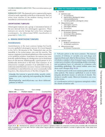
tumour originates from dental epithelium of the enamel itself i) Follicular pattern is the most common. The tumour
or its epithelial residues. Sometimes, the tumour may arise consists of follicles of variable size and shape and
from the epithelial lining of a dentigerous cyst or from basal separated from each other by fibrous tissue. The structure
layer of oral mucosa. Radiologically, typical picture is of a of follicles is similar to that of enamel organ consisting of CHAPTER 19
multilocular destruction of the bone. Rare instances of an central area of stellate cells resembling stellate reticulum,
extraosseous example, presence of an embedded tooth, or and peripheral layer of cuboidal or columnar cells
unilocular ameloblastoma can occur. Tumour with histologic resembling epithelium. The central stellate areas may
resemblance to ameloblastoma can occur occasionally in the show cystic changes (Fig. 19.11).
long bone, like adamantinoma of the tibia (Chapter 28). ii) Plexiform pattern is the next common pattern after folli-
cular pattern. The tumour epithelium is seen to form
Grossly, the tumour is greyish-white, usually solid, irregular plexiform masses or network of strands. The
sometimes cystic, replacing and expanding the affected stroma is usually scanty. Microcyst formation can occur
bone. in the stroma.
Histologically, ameloblastoma can show different iii) Acanthomatous pattern is squamous metaplasia within
patterns as follows:
the islands of tumour cells. The Oral Cavity and Salivary Glands
Figure 19.11 Ameloblastoma, follicular and plexiform patterns. Epithelial follicles are composed of central area of stellate cells and peripheral
layer of cuboidal or columnar cells. Plexiform areas show irregular plexiform masses and network of strands of epithelial cells. A few areas show
central cystic change.