
Page 555 - Textbook of Pathology, 6th Edition
P. 555
hernia in 98% of cases. The condition is diagnosed radio- 539
logically in about 5% of apparently normal asymptomatic
individuals. In symptomatic cases, especially the elderly
women, the clinical features are heartburn (retrosternal
burning sensation) and regurgitation of gastric juice into the
mouth, both of which are worsened due to heavy work, lifting
weights and excessive bending.
ETIOLOGY. The basic defect is the failure of the muscle
fibres of the diaphragm that form the margin of the
oesophageal hiatus. This occurs due to shortening of the
oesophagus which may be congenital or acquired.
Congenitally short oesophagus may be the cause of
hiatus hernia in a small proportion of cases.
More commonly, it is acquired due to secondary factors
which cause fibrous scarring of the oesophagus as follows:
a) Degeneration of muscle due to aging. Figure 20.2 Oesophageal webs and rings.
b) Increased intra-abdominal pressure such as in pregnancy,
abdominal tumours etc. Congenital diverticula occur either at the upper end of
c) Recurrent oesophageal regurgitation and spasm causing the oesophagus or at the bifurcation of trachea.
inflammation and fibrosis. Acquired diverticula may be of 2 types:
d) Increase in fatty tissue in obese people causing decreased a) Pulsion (Zenker’s) type—is seen in the region of hypo-
muscular elasticity of diaphragm. pharynx and occurs due to oesophageal obstruction such as
due to chronic oesophagitis, carcinoma etc. The mucosa and
MORPHOLOGIC FEATURES. There are 3 patterns in submucosa herniate through the weakened area or through
hiatus hernia (Fig. 20.1): defect in the muscularis propria.
i) Sliding or oesophago-gastric hernia is the most b) Traction type—occurs in the lower third of oesophagus CHAPTER 20
common, occurring in 85% of cases. The herniated part of from contraction of fibrous tissue such as from pleural
the stomach appears as supradiaphragmatic bell due to adhesions, scar tissue of healed tuberculous lesions in the
sliding up on both sides of the oesophagus. hilum, silicosis etc.
ii) Rolling or para-oesophageal hernia is seen in 10% of Complications of diverticula include obstruction, infec-
cases. This is a true hernia in which cardiac end of the tion, perforation, haemorrhage and carcinoma.
stomach rolls up para-oesophageally, producing an
intrathoracic sac. Oesophageal Webs and Rings
iii) Mixed or transitional hernia constitutes the remain- Radiological shadows in the oesophagus resembling ‘webs’
ing 5% cases in which there is combination of sliding and and ‘rings’ are observed in some patients complaining of
rolling hiatus hernia. dysphagia. The Gastrointestinal Tract
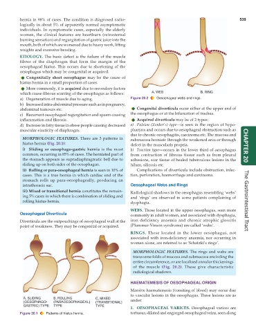
WEBS. Those located in the upper oesophagus, seen more
Oesophageal Diverticula commonly in adult women, and associated with dysphagia,
Diverticula are the outpouchings of oesophageal wall at the iron deficiency anaemia and chronic atrophic glossitis
point of weakness. They may be congenital or acquired. (Plummer-Vinson syndrome) are called ‘webs’.
RINGS. Those located in the lower oesophagus, not
associated with iron-deficiency anaemia, nor occurring in
women alone, are referred to as ‘Schatzki’s rings’.
MORPHOLOGIC FEATURES. The rings and webs are
transverse folds of mucosa and submucosa encircling the
entire circumference, or are localised annular thickenings
of the muscle (Fig. 20.2). These give characteristic
radiological shadows.
HAEMATEMESIS OF OESOPHAGEAL ORIGIN
Massive haematemesis (vomiting of blood) may occur due
to vascular lesions in the oesophagus. These lesions are as
under:
1. OESOPHAGEAL VARICES. Oesophageal varices are
Figure 20.1 Patterns of hiatus hernia. tortuous, dilated and engorged oesophageal veins, seen along