
Page 556 - Textbook of Pathology, 6th Edition
P. 556
540 the longitudinal axis of oesophagus. They occur as a result of chronic disease such as nodularity, strictures,
of elevated pressure in the portal venous system, most ulcerations and erosions.
commonly in cirrhosis of the liver (Chapter 22). Less common Microscopically, the reflux changes in the distal
causes are: portal vein thrombosis, hepatic vein thrombosis oesophagus include basal cell hyperplasia and deep
(Budd-Chiari syndrome) and pylephlebitis. The lesions occur elongation of the papillae touching close to the surface
as a result of bypassing of portal venous blood from the liver epithelium. Inflammatory changes vary according to the
to the oesophageal venous plexus. The increased venous stage of the disease. In early stage, mucosa and submucosa
pressure in the superficial veins of the oesophagus may result are infiltrated by some polymorphs and eosinophils; in
in ulceration and massive bleeding. chronic stage, there is lymphocytic infiltration and fibrosis
2. MALLORY-WEISS SYNDROME. In this condition, of all the layers of the oesophageal wall.
there is lacerations of mucosa at the gastro-oesophageal Barrett’s Oesophagus
junction following minor trauma such as by vomiting,
retching or vigorous coughing. Patients present with upper This is a condition in which, following reflux oesophagitis,
gastro-oesophageal bleeding. stratified squamous epithelium of the lower oesophagus is
replaced by columnar epithelium (columnar metaplasia). The
3. RUPTURE OF THE OESOPHAGUS. Rupture of the condition is seen more commonly in later age and is caused
oesophagus may occur following trauma, during by factors producing gastro-oesophageal reflux disease
oesophagoscopy, indirect injury (e.g. due to sudden accele- (described above). Barrett’s oesophagus is a premalignant
ration and deceleration of the body) and spontaneous rupture condition evolving sequentially from Barrett’s epithelium
(e.g. after overeating, extensive aerophagy etc). (columnar metaplasia) → dysplasia → carcinoma in situ →
4. OTHER CAUSES. Oesophageal haematemesis may also oesophageal adenocarcinoma.
occur in the following conditions:
i) Bursting of aortic aneurysm into the lumen of oesophagus MORPHOLOGIC FEATURES. Endoscopically, the
affected area is red and velvety. Hiatus hernia and peptic
ii) Vascular erosion by malignant growth in the vicinity ulcer at squamocolumnar junction (Barrett’s ulcer) are
iii) Hiatus hernia frequently associated.
iv) Oesophageal cancer Microscopically, the most common finding is the replace-
v) Purpuras ment of squamous epithelium by metaplastic columnar
vi) Haemophilia. cells. Barrett’s oesophagus may be composed of intestinal
SECTION III
epithelium, fundic gastric glands, or cardiac mucous
INFLAMMATORY LESIONS glands. Other cells present in the glands may be Paneth
cells (Fig. 20.3), goblet cells, chief cells, parietal cells,
Inflammation of the oesophagus, oesophagitis, occurs most
commonly from reflux, although a number of other clinical mucus-secreting cells and endocrine cells.
conditions and infections may also cause oesophagitis as Inflammatory changes, acute or chronic, are commonly
under: accompanied. Dysplastic changes of the columnar
epithelium or glands may be present.
Surveillance endoscopic biopsies are advised because
Reflux (Peptic) Oesophagitis
Barrett’s intestinal metaplasaia may develop dysplasia.
Reflux of the gastric juice is the commonest cause of
oesophagitis.
Systemic Pathology
PATHOGENESIS. Gastro-oesophageal reflux, to an extent,
may occur in normal healthy individuals after meals and in
early pregnancy. However, in some clinical conditions, the
gastro-oesophageal reflux is excessive, resulting in
inflammation of the lower oesophagus. These conditions are
as under:
i) Sliding hiatus hernia
ii) Chronic gastric and duodenal ulcers
iii) Nasogastric intubation
iv) Persistent vomiting
v) Surgical vagotomy
vi) Neuropathy in alcoholics, diabetics
vii) Oesophagogastrostomy.
MORPHOLOGIC FEATURES. Endoscopically, the
demarcation between normal squamous and columnar
epithelium at the junctional mucosa is lost. The affected
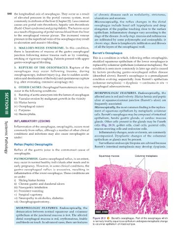
distal oesophageal mucosa is red, erythematous, friable Figure 20.3 Barrett’s oesophagus. Part of the oesophagus which
and bleeds on touch. In advanced cases, there are features is normally lined by squamous epithelium undergoes metaplastic change
to columnar epithelium of intestinal type.