
Page 557 - Textbook of Pathology, 6th Edition
P. 557
High-grade dysplasia may progress to invasive men over 50 years of age. Prognosis is dismal: with standard 541
adenocarcinoma of the oesophagus in up to 20% cases. methods of therapy (surgical resection and/or irradiation),
70% of the patients die within one year of diagnosis. Five-
Infectious Oesophagitis year survival rate is 5-10%.
A number of opportunistic infections in immunosuppressed ETIOLOGY. Although exact etiology of carcinoma of the
individuals can cause oesophagitis. Some of these agents are oesophagus is not known, a number of conditions and factors
as follows: have been implicated as under:
i) Candida (Monilial) oesophagitis
ii) Herpes simplex (Herpetic) oesophagitis 1. Diet and personal habits:
iii) Cytomegalovirus i) Heavy smoking
iv) Tuberculosis. ii) Alcohol consumption
iii) Intake of foods contaminated with fungus
Other Causes of Oesophagitis iv) Nutritional deficiency of vitamins and trace elements.
i) Eosinophilic oesophagitis caused by radiation, corrosives 2. Oesophageal disorders:
ii) Intake of certain drugs (anticholinergic drugs, i) Oesophagitis (especially Barrett’s oesophagus in
doxycycline, tetracycline) adenocarcinoma)
iii) Ingestion of hot, irritating fluids ii) Achalasia
iv) Crohn’s disease iii) Hiatus hernia
v) Various vesiculobullous skin diseases. iv) Diverticula
v) Plummer-Vinson syndrome.
TUMOURS OF OESOPHAGUS 3. Other factors:
Benign tumours of the oesophagus are uncommon and small i) Race—more common in the Chinese and Japanese than
in size (less than 3 cm). The epithelial benign tumours project in Western races; more frequent in blacks than whites.
as intraluminal masses arising from squamous epithelium ii) Family history—association with tylosis (keratosis
(squamous cell papilloma), or from columnar epithelium palmaris et plantaris). CHAPTER 20
(adenoma). The stromal or mesenchymal benign tumours are iii) Genetic factors—predisposition with coeliac disease,
intramural masses such as leiomyoma and others like lipoma, epidermolysis bullosa, tylosis.
fibroma, neurofibroma, rhabdomyoma, lymphangioma and iv) HPV infection—is the recent addition in etiologic factors.
haemangioma. At molecular level, abnormality of p53 tumour suppressor
For all practical purposes, malignant tumours of the oeso- gene has been found associated with a number of above risk
phagus are carcinomas because sarcomas such as factors, notably with consumption of tobacco and alcohol,
leiomyosarcoma and fibrosarcoma occur with extreme rarity. and in cases having proven Barrett’s oesophagus.
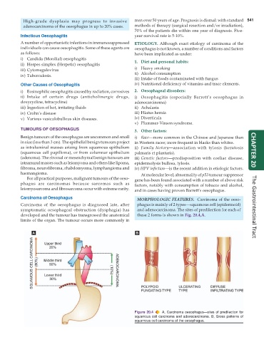
Carcinoma of Oesophagus MORPHOLOGIC FEATURES. Carcinoma of the oeso-
Carcinoma of the oesophagus is diagnosed late, after phagus is mainly of 2 types—squamous cell (epidermoid)
symptomatic oesophageal obstruction (dysphagia) has and adenocarcinoma. The sites of predilection for each of The Gastrointestinal Tract
developed and the tumour has transgressed the anatomical these 2 forms is shown in Fig. 20.4,A.
limits of the organ. The tumour occurs more commonly in
Figure 20.4 A, Carcinoma oesophagus—sites of predilection for
squamous cell carcinoma and adenocarcinoma. B, Gross patterns of
squamous cell carcinoma of the oesophagus.