
Page 558 - Textbook of Pathology, 6th Edition
P. 558
542
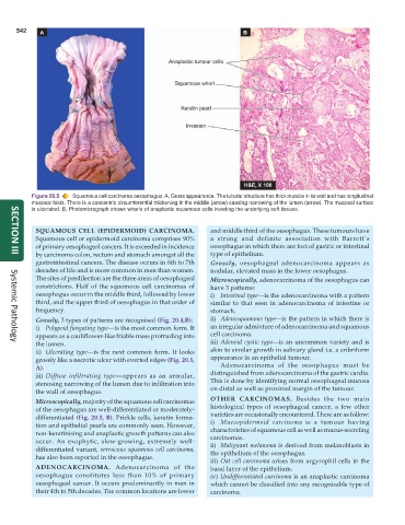
Figure 20.5 Squamous cell carcinoma oesophagus. A, Gross appearance. The tubular structure has thick muscle in its wall and has longitudinal
mucosal folds. There is a concentric circumferential thickening in the middle (arrow) causing narrowing of the lumen (arrow). The mucosal surface
is ulcerated. B, Photomicrograph shows whorls of anaplastic squamous cells invading the underlying soft tissues.
SQUAMOUS CELL (EPIDERMOID) CARCINOMA. and middle third of the oesophagus. These tumours have
Squamous cell or epidermoid carcinoma comprises 90% a strong and definite association with Barrett’s
of primary oesophageal cancers. It is exceeded in incidence oesophagus in which there are foci of gastric or intestinal
by carcinoma colon, rectum and stomach amongst all the type of epithelium.
SECTION III
gastrointestinal cancers. The disease occurs in 6th to 7th Grossly, oesophageal adenocarcinoma appears as
decades of life and is more common in men than women. nodular, elevated mass in the lower oesophagus.
The sites of predilection are the three areas of oesophageal Microscopically, adenocarcinoma of the oesophagus can
constrictions. Half of the squamous cell carcinomas of have 3 patterns:
oesophagus occur in the middle third, followed by lower i) Intestinal type—is the adenocarcinoma with a pattern
third, and the upper third of oesophagus in that order of similar to that seen in adenocarcinoma of intestine or
frequency. stomach.
Grossly, 3 types of patterns are recognised (Fig. 20.4,B): ii) Adenosquamous type—is the pattern in which there is
i) Polypoid fungating type—is the most common form. It an irregular admixture of adenocarcinoma and squamous
appears as a cauliflower-like friable mass protruding into cell carcinoma.
the lumen. iii) Adenoid cystic type—is an uncommon variety and is
Systemic Pathology
ii) Ulcerating type—is the next common form. It looks akin to similar growth in salivary gland i.e. a cribriform
grossly like a necrotic ulcer with everted edges (Fig. 20.5, appearance in an epithelial tumour.
A). Adenocarcinoma of the oesophagus must be
iii) Diffuse infiltrating type—appears as an annular, distinguished from adenocarcinoma of the gastric cardia.
stenosing narrowing of the lumen due to infiltration into This is done by identifying normal oesophageal mucosa
the wall of oesophagus. on distal as well as proximal margin of the tumour.
Microscopically, majority of the squamous cell carcinomas OTHER CARCINOMAS. Besides the two main
of the oesophagus are well-differentiated or moderately- histological types of oesophageal cancer, a few other
differentiated (Fig. 20.5, B). Prickle cells, keratin forma- varieties are occasionally encountered. These are as follow:
tion and epithelial pearls are commonly seen. However, i) Mucoepidermoid carcinoma is a tumour having
non-keratinising and anaplastic growth patterns can also characteristics of squamous cell as well as mucus-secreting
carcinomas.
occur. An exophytic, slow-growing, extremely well- ii) Malignant melanoma is derived from melanoblasts in
differentiated variant, verrucous squamous cell carcinoma, the epithelium of the oesophagus.
has also been reported in the oesophagus.
iii) Oat cell carcinoma arises from argyrophil cells in the
ADENOCARCINOMA. Adenocarcinoma of the basal layer of the epithelium.
oesophagus constitutes less than 10% of primary iv) Undifferentiated carcinoma is an anaplastic carcinoma
oesophageal cancer. It occurs predominantly in men in which cannot be classified into any recognisable type of
their 4th to 5th decades. The common locations are lower carcinoma.