
Page 559 - Textbook of Pathology, 6th Edition
P. 559
v) Carcinosarcoma consists of malignant epithelial as well is inner concavity on the right, while the greater curvature is 543
as sarcomatous components. the outer convexity on the left side of the stomach.
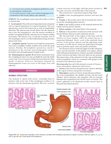
vi) Secondary tumours rarely occur in the oesophagus from The stomach has 5 anatomical regions (Fig. 20.6):
carcinomas of the breast, kidney and adrenals. 1. Cardia is the oesophagogastric junction and lacks the
sphincter.
SPREAD. The oesophageal cancer spreads locally as well as 2. Fundus is the portion above the horizontal line drawn
to distant sites. across the oesophagogastric junction.
i) Local spread. This is the most important mode of spread 3. Body is the middle portion of the stomach between the
and is of great importance for surgical treatment. The local fundus and the pyloric antrum.
spread may occur in the transverse as well as longitudinal 4. Pyloric antrum is the distal third of the stomach.
direction. The tumour may invade below into the stomach,
above into the hypopharynx, into the trachea resulting in 5. Pylorus is the junction of distal end of the stomach with
tracheo-oesophageal fistula, and may involve larynx causing the duodenum. It has powerful sphincter muscle.
hoarseness. The tumour may invade the muscular wall of The mucosal folds in the region of the body and the
the oesophagus and involve the mediastinum, lungs, bronchi, fundus are loose (rugae), while the antral mucosa is
pleura and aorta. somewhat flattened. Gastric canal is the relatively fixed
portion of the pyloric antrum and the adjoining lesser
ii) Lymphatic spread. Submucosal lymphatic permeation curvature; it is the site for numerous pathological changes
may lead to multiple satellite nodules away from the main such as gastritis, peptic ulcer and gastric carcinoma.
tumour. Besides, the lymphatic spread may result in The stomach receives its blood supply from the left gastric
metastases to the cervical, para-oesophageal, tracheo- artery and the branches of the hepatic and splenic arteries
bronchial and subdiaphragmatic lymph nodes.
with widespread anastomoses. Numerous gastric lymphatics
iii) Haematogenous spread. Blood-borne metastases from which communicate freely with each other are also present.
the oesophageal cancer are rare, probably because the death The innervation of the stomach is by the vagi and branches
occurs early due to invasion of important structures by other of the sympathetic which are connected with ganglia in the
modes of spread. However, metastatic deposits by muscular and submucous layers.
haematogenous route can occur in the lungs, liver and Histologically, the wall of the stomach consists of 4 layers— CHAPTER 20
adrenals. serosa, muscularis, submucosa and mucosa.
1. Serosa is derived from the peritoneum which is deficient
STOMACH in the region of lesser and greater curvatures.
2. Muscularis consists of 3 layers of smooth muscle fibres—
NORMAL STRUCTURE the outer longitudinal, the middle circular and the inner
oblique. Nerve plexuses and ganglion cells are present
The stomach is ‘gland with cavity’, extending from its between the longitudinal and circular layers of muscle. The
junction with lower end of the oesophagus (cardia) to its pyloric sphincter is the thickened circular muscle layer at
junction with the duodenum (pylorus). The lesser curvature the gastroduodenal junction. The Gastrointestinal Tract
Figure 20.6 Anatomical subdivisions of the stomach correlated with histological appearance of gastric mucosa in different regions. D , D , D 3
2
1
and D 4 are the first to fourth parts of the duodenum.