
Page 560 - Textbook of Pathology, 6th Edition
P. 560
544 3. Submucosa is a layer of loose fibroconnective tissue Hydrochloric acid is produced by the parietal (oxyntic) cells
binding the mucosa to the muscularis loosely and contains by the interaction of Cl’ ions of the arterial blood with water
branches of blood vessels, lymphatics and nerve plexuses and carbon dioxide in the presence of the enzyme, carbonic
and ganglion cells. anhydrase. The degree of gastric activity is correlated with
4. Mucosa consists of 2 layers—superficial and deep. the ‘total parietal cell mass’. Injection of histamine can
Between the two layers is the lamina propria composed of stimulate the production of acid component of the gastric
network of fibrocollagenic tissue with a few lymphocytes, juice, while the pepsin-secreting chief cells do not respond
plasma cells, macrophages and eosinophils. The mucosa is to histamine. Physiologically, the gastric secretions are
externally bounded by muscularis mucosae: stimulated by the food itself.
i) Superficial layer. It consists of a single layer of surface The control of gastric secretions chiefly occurs in one of the
epithelium composed of regular, mucin-secreting, tall following 3 ways:
columnar cells with basal nuclei. There is a very rapid 1. Cephalic phase—is stimulated by the sight, smell, taste
turnover of these cells. These dip down at places to form or even thought of food. A neural reflex is initiated via
crypts (or pits or foveolae). branches of the vagus nerve that promotes the release of
Cardiac mucosa is the transition zone between the oeso- hydrochloric acid, pepsinogen and mucus.
phageal squamous mucosa and the oxyntic mucosa of the 2. Gastric phase—is triggered by the mechanical and
fundus and body with which it gradually merges. chemical stimuli.
Oxyntic mucosa lines both gastric fundus and body. i) Mechanical stimulation comes from stretching of the wall
Antral mucosa lines the pyloric antrum. of the stomach and conveying neural messages to the medulla
ii) Deep layer: It consists of glands that open into the for gastric secretion.
bottom of the crypts. Depending upon the structure, these ii) Chemical stimulation is by digested proteins, amino acids,
glands are of 3 types: bile salts and alcohol which act on gastrin-producing G cells.
a) Glands of the cardia are simple tubular or compound Gastrin then passes into the blood stream and on return to
tubulo-racemose, lined by mucin secreting cells. A few the stomach promotes the release of gastric juice.
endocrine cells and occasional parietal and chief cells are also 3. Intestinal phase—is triggered by the entry of protein-
present. rich food in the small intestine. An intestinal hormone capable
b) Glands of the body-fundus are long, tubular and tightly of stimulating gastric secretion is probably released into the
packed which may be coiled or dilated. There are 4 types of blood stream.
cells present in the glands of body-fundic mucosa:
SECTION III
Parietal (Oxyntic) cells—are the most numerous and line GASTRIC ANALYSIS
the superficial (upper) part of the glands. Parietal cells In various diseases of the stomach, the laboratory tests to
are triangular in shape, have dark-staining nuclei and measure gastric secretions (consisting of gastric acid, pepsin,
eosinophilic cytoplasm. These cells are responsible for mucus and intrinsic factor) and serum gastrin are of
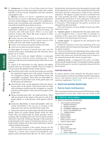
production of hydrochloric acid of the gastric juice and particular significance (Table 20.1).
the blood group substances.
Chief (Peptic) cells—are the dominant cells in the deeper A. TESTS FOR GASTRIC SECRETIONS
(lower) parts of the glands. Their basal nuclei are large
with prominent nucleoli and the cytoplasm is coarsely 1. Tests for Gastric Acid Secretions
granular and basophilic. These cells secrete pepsin of the The conventional fractional test meal (FTM) has been totally
gastric juice. superseded by newer tests. These tests are based on the
Systemic Pathology
Mucin-secreting neck cells—are small and fewer. These
cells are present in the region of the narrow neck of the TABLE 20.1: Gastric Analysis.
gastric glands i.e. at the junction of the glands with the
pits. A. TESTS FOR GASTRIC SECRETIONS
1. Tests for gastric acid secretions
Endocrine (Kulchitsky or Enterochromaffin) cells—are i) Histamine stimulation
widely distributed in the mucosa of all parts of the ii) Histalog stimulation
alimentary tract and are described later (page 561). iii) Pentagastrin (peptavlon) stimulation
c) Glands of the pylorus are much longer than the body-fundic iv) Insulin meal (Hollander test)
glands. These are simple tubular glands which are often v) Tubeless analysis
coiled. They are lined mainly by small, granular, mucin- 2. Tests for pepsin
secreting cells resembling neck cells and occasional parietal Pepsin inhibitors
cells but no chief cells. Gastrin-producing G-cells are present 3. Tests for mucus
Protein content of mucus
predominantly in the region of antropyloric mucosa, with a 4. Tests for intrinsic factor
small number of these cells in the crypts and Brunner’s glands
of the proximal duodenum. B. TESTS FOR GASTRIN
The secretory products of the gastric mucosa are the 1. Serum gastrin
gastric juice and the intrinsic factor, required for absorption of 2. Gastrin provocation tests
vitamin B . Gastric juice consists of hydrochloric acid, ii) i) Secretin test
Calcium infusion test
12
–
+
+
pepsin, mucin and electrolytes like Na , K , HCO’ and Cl .
3