
Page 564 - Textbook of Pathology, 6th Edition
P. 564
548
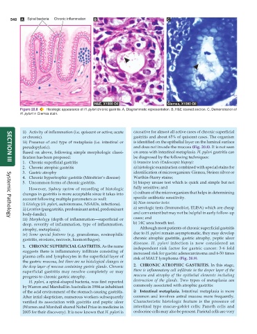
Figure 20.8 Histologic appearance of H. pylori chronic gastritis. A, Diagrammatic representation. B, H&E stained section. C, Demonstration of
H. pylori in Giemsa stain.
ii) Activity of inflammation (i.e. quiscent or active; acute causative for almost all active cases of chronic superficial
or chronic). gastritis and about 65% of quiscent cases. The organism
iii) Presence of and type of metaplasia (i.e. intestinal or is identified on the epithelial layer on the luminal surface
pseudopyloric). and does not invade the mucosa (Fig. 20.8). It is not seen
Based on above, following simple morphologic classi- on areas with intestinal metaplasia. H. pylori gastritis can
fication has been proposed: be diagnosed by the following techniques:
1. Chronic superficial gastritis i) Invasive tests (Endoscopic biopsy):
SECTION III
2. Chronic atrophic gastritis a) histologic examination combined with special stains for
3. Gastric atrophy identification of microorganism: Giemsa, Steiner silver or
4. Chronic hypertrophic gastritis (Ménétrier’s disease) Warthin-Starry stains;
5. Uncommon forms of chronic gastritis. b) biopsy urease test which is quick and simple but not
However, Sydney system of recording of histologic fully sensitive; and
changes in gastritis is more acceptable since it takes into c) culture of the microorganism that helps in determining
account following multiple parameters as well: specific antibiotic sensitivity.
i) Etiology (H. pylori, autoimmune, NSAIDs, infections). ii) Non-invasive tests:
ii) Location (pangastritis, predominant antral, predominant a) serologic tests (Immunoblot, ELISA) which are cheap
body-fundic). and convenient but may not be helpful in early follow-up
iii) Morphology (depth of inflammation—superficial or cases; and
Systemic Pathology
deep, severity of inflammation, type of inflammation, b) 14C urea breath test.
atrophy, metaplasia). Although most patients of chronic superficial gastritis
iv) Some special features (e.g. granulomas, eosinophilic due to H. pylori remain asymptomatic, they may develop
gastritis, erosions, necrosis, haemorrhages). chronic atrophic gastritis, gastric atrophy, peptic ulcer
disease. H. pylori infection is now considered an
1. CHRONIC SUPERFICIAL GASTRITIS. As the name independent risk factor for gastric cancer: 3-6 fold
suggests there is inflammatory infiltrate consisting of increased risk for gastric adenocarcinoma and 6-50 times
plasma cells and lymphocytes in the superficial layer of risk of MALT lymphoma (Fig. 20.9).
the gastric mucosa, but there are no histological changes in
the deep layer of mucosa containing gastric glands. Chronic 2. CHRONIC ATROPHIC GASTRITIS. In this stage,
superficial gastritis may resolve completely or may there is inflammatory cell infiltrate in the deeper layer of the
progress to chronic gastric atrophy. mucosa and atrophy of the epithelial elements including
H. pylori, a spiral-shaped bacteria, was first reported destruction of the glands. Two types of metaplasia are
by Warren and Marshall in Australia in 1984 as inhabitant commonly associated with atrophic gastritis:
of the acid environment of the stomach causing gastritis. i) Intestinal metaplasia. Intestinal metaplasia is more
After intial skepticism, numerous workers subsequently common and involves antral mucosa more frequently.
verified its association with gastritis and peptic ulcer Characteristic histologic feature is the presence of
(Warren and Marshall shared Nobel Prize in medicine in intestinal type mucus-goblet cells; Paneth cells and
2005 for their discovery). It is now known that H. pylori is endocrine cells may also be present. Parietal cells are very