
Page 565 - Textbook of Pathology, 6th Edition
P. 565
muscularis mucosae may extend into the thickened folds. 549
Epithelium-lined cysts are commonly seen in the
glandular layer. Inflammatory infiltrate is usually mild
but lymphoid follicles may be present. The condition is
considered significant in view of the risk of developing
cancer.
5. MISCELLANEOUS FORMS OF CHRONIC GAS-
TRITIS. A few other types of gastritis which do not fit
into the description of the types of gastritis described
above are as under:
i) Eosinophilic gastritis. This condition is characterised
by diffuse thickening of the pyloric antrum due to oedema
and extensive infiltration by eosinophils in all the layers
of the wall of antrum. Eosinophilic gastritis probably has
Figure 20.9 Consequences of long-term H. pylori gastritis. an allergic basis.
iii) Chronic follicular gastritis. This is a variant of chronic
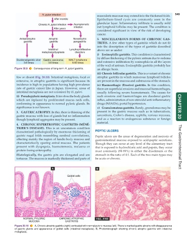
few or absent (Fig. 20.10). Intestinal metaplasia, focal or atrophic gastritis in which numerous lymphoid follicles
extensive, in atrophic gastritis is significant because its are present in the mucosa and submucosa of the stomach.
incidence is high in populations having high prevalence iv) Haemorrhagic (Erosive) gastritis. In this condition,
rate of gastric cancer like in Japan. However, areas of there are superficial erosions and mucosal haemorrhages,
intestinal metaplasia are not colonised by H. pylori. usually following severe haematemesis. The causes for
ii) Pseudopyloric metaplasia. It involves the body glands such erosions and haemorrhages are duodenal-gastric
which are replaced by proliferated mucus neck cells, reflux, administration of non-steroidal anti-inflammatory
conforming in appearance to normal pyloric glands. Its drugs (NSAIDs), portal hypertension.
significance is not known. v) Granulomatous gastritis. Rarely, granulomas may be CHAPTER 20
3. GASTRIC ATROPHY. In this, there is thinning of the present in the gastric mucosa such as in tuberculosis,
gastric mucosa with loss of glands but no inflammation sarcoidosis, Crohn’s disease, syphilis, various mycoses,
though lymphoid aggregates may be present. and as a reaction to endogenous substance or foreign
material.
4. CHRONIC HYPERTROPHIC GASTRITIS (MÉNÉ-
TRIER’S DISEASE). This is an uncommon condition
characterised pathologically by enormous thickening of PEPTIC ULCERS
gastric rugal folds resembling cerebral convolutions, Peptic ulcers are the areas of degeneration and necrosis of
affecting mainly the region of fundic-body mucosa and gastrointestinal mucosa exposed to acid-peptic secretions.
characteristically sparing antral mucosa. The patients Though they can occur at any level of the alimentary tract
present with dyspepsia, haematemesis, melaena or that is exposed to hydrochloric acid and pepsin, they occur The Gastrointestinal Tract
protein-losing enteropathy. most commonly (98-99%) in either the duodenum or the
Histologically, the gastric pits are elongated and are stomach in the ratio of 4:1. Each of the two main types may
tortuous. The mucosa is markedly thickened and parts of be acute or chronic.
Figure 20.10 A, Chronic atrophic gastritis (right) contrasted with normal pyloric mucosa (left). There is marked gastric atrophy with disappearance
of gastric glands and appearance of goblet cells (intestinal metaplasia). B, Photomicrograph showing chronic atrophic gastritis with intestinal
metaplasia.