
Page 568 - Textbook of Pathology, 6th Edition
P. 568
552 1. There is generally hypersecretion of gastric acid into the
fasting stomach at night which takes place under the
influence of vagal stimulation. There is high basal as well as
maximal acid output (BAO and MAO) in response to various
stimuli.
2. Patients of duodenal ulcer have rapid emptying of the
stomach so that the food which normally buffers and
neutralises the gastric acid, passes down into the small
intestine, leaving the duodenal mucosa exposed to the
aggressive action of gastric acid.
3. Helicobacter gastritis caused by H. pylori is seen in 95-100%
cases of duodenal ulcers. The underlying mechanisms are
as under:
i) Gastric mucosal defense is broken by bacterial elaboration
of urease, protease, catalase and phospholipase.
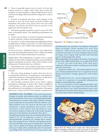
ii) Host factors: H. pylori-infected mucosal epithelium releases Figure 20.11 Distribution of peptic ulcers.
proinflammatory cytokines such as IL-1, IL-6, IL-8 and tumour
necrosis factor-α, all of which incite intense inflammatory duodenal ulcers are coexistent. Vast majority of the peptic
reaction. ulcers are benign. Chronic duodenal ulcer never turns
iii) Bacterial factors: Epithelial injury is also induced by malignant, while chronic gastric ulcer may develop
cytotoxin-associated gene protein (CagA), while vacuolating carcinoma in less than 1% of cases. Malignant gastric ulcers
cytotoxin (VacA) induces elaboration of cytokines. are larger, bowl-shaped with elevated and indurated
mucosa at the margin (Fig. 20.13).
Gastric ulcer. The pathogenesis of gastric ulcer is mainly
explained on the basis of impaired gastric mucosal defenses Microscopically, chronic peptic ulcers have 4 histological
against acid-pepsin secretions. Some other features in the zones. From within outside, these are as under (Fig. 20.14):
pathogenesis of gastric ulcer are as follows: 1. Necrotic zone—lies in the floor of the ulcer and is
composed of fibrinous exudate containing necrotic debris
1. Hyperacidity may occur in gastric ulcer due to increased and a few leucocytes.
SECTION III
serum gastrin levels in response to ingested food in an atonic 2. Superficial exudative zone—lies underneath the necrotic
stomach. zone. The tissue elements here show coagulative necrosis
2 However, many patients of gastric ulcer have low-to- giving eosinophilic, smudgy appearance with nuclear
normal gastric acid levels. Ulcerogenesis in such patients is debris.
explained on the basis of damaging influence of other factors 3. Granulation tissue zone—is seen merging into the necro-
such as gastritis, bile reflux, cigarette smoke etc. tic zone. It is composed of nonspecific inflammatory
3. The normally protective gastric mucus ‘barrier’ against infiltrate and proliferating capillaries.
acid-pepsin is deranged in gastric ulcer. There is depletion 4. Zone of cicatrisation—is seen merging into thick layer
in the quantity as well as quality of gastric mucus. One of of granulation tissue. It is composed of dense fibrocolla-
the mechanisms for its depletion is colonisation of the gastric genic scar tissue over which granulation tissue rests.
mucosa by H. pylori seen in 75-80% patients of gastric ulcer. Thrombosed or sclerotic arteries may cross the ulcer which
Systemic Pathology
on erosion may result in haemorrhage.
MORPHOLOGIC FEATURES. Gross and microscopic
changes in gastric and duodenal ulcers are similar and
quite characteristic. Gastric ulcers are found predominantly
along the lesser curvature in the region of pyloric antrum,
more commonly on the posterior than the anterior wall.
Most duodenal ulcers are found in the first part of the
duodenum, usually immediate post-pyloric, more
commonly on the anterior than the posterior wall.
Uncommon locations include ulcer in the cardia, marginal
ulcer and in the Meckel’s diverticulum (Fig. 20.11).
Grossly, typical peptic ulcers are commonly solitary (80%),
small (1-2.5 cm in diameter), round to oval and
characteristically ‘punched out’. Benign ulcers usually
have flat margins in level with the surrounding mucosa.
The mucosal folds converge towards the ulcer. The ulcers
may vary in depth from being superficial (confined to
mucosa) to deep ulcers (penetrating into the muscular Figure 20.12 Benign chronic peptic ulcer. Partial gastrectomy
layer) (Fig. 20.12). In about 10-20% of cases, gastric and specimen showing a punched out round to oval ulcer on the mucosa,
about 1 cm in diameter (arrow) and penetrating into muscularis layer.