
Page 569 - Textbook of Pathology, 6th Edition
P. 569
i) On perforation the contents escape into the lesser sac or 553
into the peritoneal cavity, causing acute peritonitis.
ii) Air escapes from the stomach and lies between the liver
and the diaphragm giving the characteristic radiological
appearance of air under the diaphragm.
iii) Subphrenic abscess between the liver and the diaphragm
may develop due to infection.
iv) Perforation may extend to involve the adjacent organs e.g.
the liver and pancreas.
4. Malignant transformation. The dictum ‘cancers ulcerate
but ulcers rarely cancerate’ holds true for most peptic ulcers.
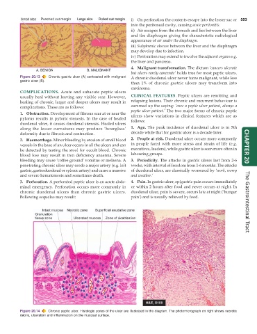
Figure 20.13 Chronic gastric ulcer (A) contrasted with malignant A chronic duodenal ulcer never turns malignant, while less
gastric ulcer (B).
than 1% of chronic gastric ulcers may transform into
carcinoma.
COMPLICATIONS. Acute and subacute peptic ulcers
usually heal without leaving any visible scar. However, CLINICAL FEATURES. Peptic ulcers are remitting and
healing of chronic, larger and deeper ulcers may result in relapsing lesions. Their chronic and recurrent behaviour is
complications. These are as follows: summed up the saying: ‘once a peptic ulcer patient, always a
peptic ulcer patient.’ The two major forms of chronic peptic
1. Obstruction. Development of fibrous scar at or near the ulcers show variations in clinical features which are as
pylorus results in pyloric stenosis. In the case of healed follows:
duodenal ulcer, it causes duodenal stenosis. Healed ulcers
along the lesser curvatures may produce ‘hourglass’ 1. Age. The peak incidence of duodenal ulcer is in 5th
deformity due to fibrosis and contraction. decade while that for gastric ulcer is a decade later.
2. Haemorrhage. Minor bleeding by erosion of small blood 2. People at risk. Duodenal ulcer occurs more commonly
vessels in the base of an ulcer occurs in all the ulcers and can in people faced with more stress and strain of life (e.g. CHAPTER 20
be detected by testing the stool for occult blood. Chronic executives, leaders), while gastric ulcer is seen more often in
blood loss may result in iron deficiency anaemia. Severe labouring groups.
bleeding may cause ‘coffee ground’ vomitus or melaena. A 3. Periodicity. The attacks in gastric ulcers last from 2-6
penetrating chronic ulcer may erode a major artery (e.g. left weeks, with interval of freedom from 1-6 months. The attacks
gastric, gastroduodenal or splenic artery) and cause a massive of duodenal ulcer, are classically worsened by ‘work, worry
and severe hematemesis and sometimes death. and weather.’
3. Perforation. A perforated peptic ulcer is an acute abdo- 4. Pain. In gastric ulcer, epigastric pain occurs immediately
minal emergency. Perforation occurs more commonly in or within 2 hours after food and never occurs at night. In
chronic duodenal ulcers than chronic gastric ulcers. duodenal ulcer, pain is severe, occurs late at night (‘hunger
Following sequelae may result: pain’) and is usually relieved by food. The Gastrointestinal Tract
Figure 20.14 Chronic peptic ulcer. Histologic zones of the ulcer are illustrated in the diagram. The photomicrograph on right shows necrotic
debris, ulceration and inflammation on the mucosal surface.