
Page 570 - Textbook of Pathology, 6th Edition
P. 570
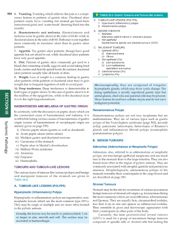
554 5. Vomiting. Vomiting which relieves the pain is a conspi- TABLE 20.4: Gastric Tumours and Tumour-like Lesions.
cuous feature in patients of gastric ulcer. Duodenal ulcer
patients rarely have vomiting but instead get heart-burn A. TUMOUR-LIKE LESIONS (POLYPS)
(retrosternal pain) and ‘water brash’ (burning fluid into the 1. Hyperplastic (inflammatory) polyps
2. Hamartomatous polyps
mouth).
B. BENIGN TUMOURS
6. Haematemesis and melaena. Haematemesis and 1. Epithelial
melaena occur in gastric ulcers in the ratio of 60:40, while in Adenomas (adenomatous or neoplastic polyps)
duodenal ulcers in the ratio of 40:60. Both may occur together 2. Non-epithelial
more commonly in duodenal ulcer than in gastric ulcer Gastrointestinal spindle cell (stromal) tumours (GIST)
patients. C. MALIGNANT TUMOURS
7. Appetite. The gastric ulcer patients, though have good 1. Epithelial (90%)
appetite but are afraid to eat, while duodenal ulcer patients (i) Adenocarcinoma
have very good appetite. (ii) Others
2. Non-epithelial (2%)
8. Diet. Patients of gastric ulcer commonly get used to a (i) Leiomyosarcoma
bland diet consisting of milk, eggs etc and avoid taking fried (ii) Leiomyoblastoma
foods, curries and heavily spiced foods. In contrast, duodenal (epithelioid leiomyoma)
ulcer patients usually take all kinds of diets. 3. Carcinoid tumour (3%)
4. Lymphoma (4%)
9. Weight. Loss of weight is a common finding in gastric
ulcer patients while patients of duodenal ulcer tend to gain
weight due to frequent ingestion of milk to avoid pain. Microscopically, they are composed of irregular
10. Deep tenderness. Deep tenderness is demonstrable in hyperplastic glands, which may show cystic change. The
both types of peptic ulcers. In the case of gastric ulcer it is in lining epithelium is mostly superficial gastric type but
the midline of the epigastrium, while in the duodenal ulcer antral glands, chief cells and parietal cells may be present.
it is in the right hypochondrium. These lesions do not have cellular atypia and do not have
malignant potential.
HAEMATEMESIS AND MELAENA OF GASTRIC ORIGIN
Hamartomatous Polyps
In continuity with the discussion on peptic ulcers which are
the commonest cause of haematemesis and melaena, it is Hamartomatous polyps are not true neoplasms but are
SECTION III
worthwhile listing various causes of haematemesis of gastric malformations. They are of various types such as gastric
origin (causes of haematemesis of oesophageal origin are polyps of the Peutz-Jeghers syndrome (page 582), juvenile
already given on page 539). polyp, pancreatic heterotopia, heterotopia of Brunner’s
i) Chronic peptic ulcers (gastric as well as duodenal) glands and inflammatory fibroid polyps (eosinophilic
ii) Acute peptic ulcers (stress ulcers) granulomatous polyps).
iii) Multiple gastric and duodenal erosions
iv) Carcinoma of the stomach B. BENIGN TUMOURS
v) Peptic ulcer in Meckel’s diverticulum Adenomas (Adenomatous or Neoplastic Polyps)
vi) Mallory-Weiss syndrome
vii) Anaemias Adenomas, also, referred to as adenomatous or neoplastic
Systemic Pathology
viii) Purpuras polyps, are true benign epithelial neoplasms and are much
ix) Haemophilia. rare in the stomach than in the large intestine. They are also
found more often in the region of pyloric antrum. They are
TUMOURS AND TUMOUR-LIKE LESIONS commonly associated with atrophic gastritis and pernicious
anaemia. Morphologically, adenomatous polyps of the
The various types of tumour-like lesions (polyps) and benign stomach resemble their counterparts in the large bowel and
and malignant tumours of the stomach are given in are described on page 583.
Table 20.4.
Stromal Tumours
A. TUMOUR-LIKE LESIONS (POLYPS)
Stomach may be the site for occurrence of various uncommon
Hyperplastic (Inflammatory) Polyps benign tumours of stromal cell origin e.g. leiomyomas (being
Hyperplastic or inflammatory polyps are regenerative, non- the most common); others are neurofibromas, schwannomas
neoplastic lesions which are the most common type (90%). and lipomas. They are usually firm, circumscribed nodules,
They may be single or multiple and are more often located less than 4 cm in size and appear as submucosal nodules.
in the pyloric antrum. They resemble in gross and microscopic appearance with
their counterparts in other parts of the body.
Grossly, the lesions may be sessile or pedunculated, 1 cm Currently, the term gastrointestinal stromal tumours
or larger in size, smooth and soft. The surface may be (GISTs) is used for a group of uncommon benign tumours
ulcerated or haemorrhagic. composed of spindle cells or stromal cells but lacking the