
Page 573 - Textbook of Pathology, 6th Edition
P. 573
large intestine. It is seen more often in the fundus. The SPREAD. Carcinoma of the stomach may spread by the 557
tumour undergoes necrosis and infection commonly. following routes:
Histologically, fungating or polypoid carcinomas are 1. Direct spread. Direct spread by local extension is the most
well-differentiated adenocarcinomas, commonly papillary common feature of gastric carcinoma. The spread occurs
type. mainly from the loose submucosal layer but eventually
muscularis and serosa are also invaded. After the peritoneal
iii) Scirrhous carcinoma (Linitis plastica) (Fig. 20.18,D).
In this pattern, the stomach wall is thickened due to covering of the stomach has been invaded, transcoelomic
extensive desmoplasia giving the appearance as ‘leather- dissemination may occur in any other part of the peritoneal
bottle stomach’ or ‘linitis plastica’. The involvement may cavity but ovarian masses (one sided or both-sided) occur
be localised to pyloric antrum, or diffuse affecting whole more commonly, referred to as Krukenberg tumours (Chapter
of the stomach from the cardia to pylorus. The lumen of 24). Submucosal spread occurs more often upwards into the
the stomach is reduced. There are no ulcers but rugae are oesophagus due to continuity of the layers of stomach with
prominent (Fig. 20.19,C). those of oesophagus, while the spread downwards into the
Histologically, it may be an adenocarcinoma or signet- duodenum occurs less often due to the presence of pyloric
ring cell carcinoma, extensively infiltrating the stomach sphincter and submucosal Brunner’s glands. The tumour
wall, but due to marked desmoplasia cancer cells may be may directly involve other neighbouring structures and
difficult to find (Fig. 20.19,D). organs like lesser and greater omentum, pancreas, liver,
common bile duct, diaphragm, spleen and transverse colon.
iv) Colloid (Mucoid) carcinoma (Fig. 20.18,E). This 2. Lymphatic spread. Metastases to regional lymph nodes
pattern is usually seen in the fundus. The tumour grows occur early, especially in the scirrhous carcinoma. The groups
like masses having gelatinous appearance due to secretion of lymph nodes involved are along the lesser and greater
of large quantities of mucus. curvature around the cardia and suprapancreatic lymph
Histologically, mucoid carcinoma contains abundant nodes. Involvement of left supraclavicular lymph node,
pools of mucin in which are seen a small number of Virchow or Troisier’s sign, is sometimes the presenting feature
tumour cells, sometimes having signet-ring appearance.
of gastric carcinoma.
v) Ulcer-cancer (Fig. 20.18,F). Development of cancer in 3. Haematogenous spread. Blood spread of gastric CHAPTER 20
chronic gastric ulcer is a rare occurrence (less than 1%). carcinoma may occur to the liver, lungs, brain, bones, kidneys
Majority of ulcer-cancers are malignant lesions from the and adrenals. It occurs more commonly with the poorly-
beginning. For confirmation of cancer in a pre-existing differentiated carcinoma.
gastric ulcer, the characteristic microscopic appearance of The American Joint Committee on Cancer has developed
peptic ulcer should be demonstrable with one portion of TNM staging system for gastric carcinoma based on tumour
the base or the margin of the ulcer showing carcinomatous invasion (T), lymph node involvement (N) and distant
changes. metastasis (M) into earliest stage T N M (intraepithelial
0
Histologically, ulcer-cancers are adenocarcinomas tumour) to most advanced stage T is N 0 M .
without any specific features. The differences between a any any 1
benign and malignant gastric ulcer are summarised in CLINICAL FEATURES. Gastric carcinoma may have diverse
Table 20.5 (also see Fig. 20.13). presentations. The usual clinical features are as under: The Gastrointestinal Tract
i) Persistent abdominal pain
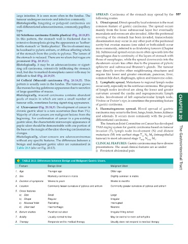
TABLE 20.5: Differences between Benign and Malignant Gastric Ulcers.
Feature Benign Ulcer Malignant Ulcer
1. Age Younger age Older age
2. Sex Markedly common in males Slightly common in males
3. Duration of symptoms Weeks to years Weeks to months
4. Location Commonly lesser curvature of pylorus and antrum Commonly greater curvature of pylorus and antrum
5. Gross features
a) Size Small Large
b) Shape Regular Irregular
c) Mucosal folds Radiating Interrupted
d) Ulcer bed Haemorrhagic Necrotic
6. Barium studies Punched out ulcer Irregular filling defect
7. Acidity Usually normal-to-low May be normal-to-even achlorhydria
8. Therapy Responds well to medical therapy Usually does not respond to medical therapy