
Page 576 - Textbook of Pathology, 6th Edition
P. 576
560
SECTION III
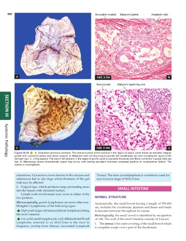
Figure 20.19 A, Ulcerative carcinoma stomach. The luminal surface of the stomach in the region of pyloric canal shows an elevated irregular
growth with ulcerated surface and raised margins. B, Malignant cells forming irregular glands with stratification are seen invading the layers of the
Systemic Pathology
stomach wall. C, Linitis plastica. The wall of the stomach in the region of pyloric canal is markedly thickened and fibrotic while the mucosal folds are
lost. D, Microscopy shows characteristic signet ring tumour cells having abundant mucinous cytoplasm positive for mucicarmine (inbox). The
stroma is desmoplastic.
ulcerations. Cut section shows lesions in the mucosa and Tissue). The term pseudolymphoma is sometimes used for
submucosa but in late stage whole thickness of the gut non-invasive stage of MALToma.
wall may be affected.
2. Polypoid type, which produces large protruding mass
into the lumen with ulcerated surface. SMALL INTESTINE
Lymph node involvement may occur in either of the
two patterns. NORMAL STRUCTURE
Microscopically, gastric lymphomas are most often non- Anatomically, the small bowel having a length of 550-650
Hodgkin’s lymphomas of the following types: cm, includes the duodenum, jejunum and ileum and tends
High-grade large cell immunoblastic lymphoma being to become narrower throughout its course.
the most common. Histologically, the small bowel is identified by recognition
Low-grade small lymphocytic well-differentiated B-cell of villi. The wall of the small intestine consists of 4 layers:
lymphoma referred to as MALToma is the next in 1. The serosa is the outer covering of the small bowel which
frequency (arising from Mucosa Associated Lymphoid is complete except over a part of the duodenum.