
Page 580 - Textbook of Pathology, 6th Edition
P. 580
564
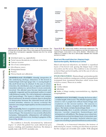
Figure 20.23 Haemorrhagic infarct of the small intestine. The Figure 20.24 Infarct small intestine, microscopic appearance. The
infarcted area is swollen, dark in colour and coated with fibrinous exudate. mucosa in the infarcted area shows coagulative necrosis and submucosal
A sharp line of demarcation separates infarcted area from the normal haemorrhages: muscularis is also partly affected. Inflammatory cell
bowel (arrow). infiltration is marked at the line of demarcation between the infarcted
and normal bowel.
Intestinal sepsis e.g. appendicitis
Portal venous thrombosis in cirrhosis of the liver Mural and Mucosal Infarction (Haemorrhagic
Tumour invasion Gastroenteropathy, Membranous Colitis)
Use of oral contraceptives Mural and mucosal infarctions are limited to superficial
iv) Miscellaneous causes: layers of the bowel wall, sparing the deeper layer of the
SECTION III
Strangulated hernia muscularis and the serosa. The condition is also referred to
Torsion as haemorrhagic gastroenteropathy, and in the case of colon as
Fibrous bands and adhesions. membranous colitis.
ETIOPATHOGENESIS. Haemorrhagic gastroenteropathy
MORPHOLOGIC FEATURES. Grossly, irrespective of results from conditions causing non-occlusive hypoperfusion
the underlying etiology, infarction of the bowel is (compared from transmural infarction which occurs from
haemorrhagic (red) type (page 126). A varying length of occlusive causes). These are as under:
the small bowel may be affected. In the case of colonic Shock
infarction, the distribution area of superior and inferior Cardiac failure
mesenteric arteries (i.e. splenic flexure) is more commonly Infections
involved. The affected areas become dark purple and
Systemic Pathology
markedly congested and the peritoneal surface is coated Intake of drugs causing vasoconstriction e.g. digitalis,
with fibrinous exudate. The wall is thickened, oedematous norepinephrine.
and haemorrhagic. The lumen is dilated and contains
blood and mucus. In arterial occlusion, there is sharp line MORPHOLOGIC FEATURES. Grossly, the lesions affect
of demarcation between the infarcted bowel and the variable length of the bowel. The affected segment of the
normal intestine, whereas in venous occlusion the bowel is red or purple but without haemorrhage and
infarcted area merges imperceptibly into the normal bowel exudation on the serosal surface. The mucosa is
(Fig. 20.23). oedematous at places, sloughed and ulcerated at other
Microscopically, there is coagulative necrosis and ulcera- places. The lumen contains haemorrhagic fluid.
tion of the mucosa and there are extensive submucosal Microscopically, there is patchy ischaemic necrosis of
haemorrhages. The muscularis is less severely affected by mucosa, vascular congestion, haemorrhages and
ischaemia. Subsequently, inflammatory cell infiltration inflammatory cell infiltrate. The changes may extend into
and secondary infection occur, leading to gangrene of the superficial muscularis but deeper layer of muscularis and
bowel (Fig. 20.24). serosa are spared. Secondary bacterial infection may
supervene resulting in pseudomembranous enterocolitis.
The condition is clinically characterised by ‘abdominal
angina’ in which the patient has acute abdominal pain, Clinically, as in transmural infarction, the features of
nausea, vomiting, and sometimes diarrhoea. The disease is abdominal pain, nausea, vomiting and diarrhoea are present,
rapidly fatal, with 50-70% mortality rate. but the changes are reversible and curable. With adequate