
Page 582 - Textbook of Pathology, 6th Edition
P. 582
566 antigens and commensal flora in the intestinal lumen. The peripheral blood. These cells either activate other
mechanism responsible for this is by activation of CD4+ T inflammatory cells (e.g. macrophages and B cells), or recruit
cells secreting cytokines inhibitory to inflammation (IL-10, more inflammatory cells by stimulation of homing receptor
TGF-β) which suppress inflammation in the gut wall. In IBD, on leucocytes and vascular endothelium. There are two main
this immune mechanism of suppression of inflammation is types of CD4+ T cells in IBD:
defective and thus results in uncontrolled inflammation. TH1 cells secrete proinflammatory cytokines IFN-γ and
ii) Transgenic mouse experimental model studies. Gene ‘knock TNF which induce transmural granulomatous inflammation
out’ studies on colitis in mice have revealed that multiple seen in Crohn’s disease. IL-12 initiates TH1 cytokine
immune abnormalities may be responsible for IBD as under: pathway.
a) Deletion of inflammation inhibitory cytokines (e.g. IL-2, TH2 cells secrete IL-4, IL-5 and IL-13 which induce
IL-10, TGF-β) or their receptors. superficial mucosal inflammation characteristically seen in
ulcerative colitis.
b) Deletion of molecules responsible for T cell recognition
(e.g. T cell antigen receptors, MHC class II). 3. Exogenous factors. In addition to role of genetic factors
and deranged T-cell mediated immunity, a role for several
c) Interference with normal epithelial barrier function in the exogenous and environmental factors has been assigned:
intestine (e.g. blocking N-cadherin, deletion of multi-drug i) Microbial factors: At different times, role of a variety of
resistance MDR gene). microbes in initiation of inflammatory response by the body
iii) Type of inflammatory cells. In both types of IBD, activated has been suspected. Accordingly, several microorganism
CD4+ T cells are present in the lamina propria and in the species (bacteria, viruses, protozoa and fungi) have been
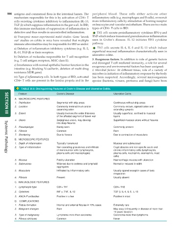
TABLE 20.6: Distinguishing Features of Crohn’s Disease and Ulcerative Colitis.
Feature Crohn’s Disease Ulcerative Colitis
A. MACROSCOPIC FEATURES
1. Distribution Segmental with skip areas Continuous without skip areas
2. Location Commonly terminal ileum and/or Commonly rectum, sigmoid colon and
ascending colon extending upwards
3. Extent Usually involves the entire thickness Usually superficial, confined to mucosal
SECTION III
of the affected segment of bowel wall layers
4. Ulcers Serpiginous ulcers, may develop Superficial mucosal ulcers without fissures
into deep fissures
5. Pseudopolyps Rarely seen Commonly present
6. Fibrosis Common Rare
7. Shortening Due to fibrosis Due to contraction of muscularis
B. MICROSCOPIC FEATURES
1. Depth of inflammation Typically transmural Mucosal and submucosal
2. Type of inflammation Non-caseating granulomas and infiltrate Crypt abscess and non-specific acute and
of mononuclear cells (lymphocytes, chronic inflammatory cells (lymphocytes,
plasma cells and macrophages) plasma cells, neutrophils, eosinophils, mast
Systemic Pathology
cells)
3. Mucosa Patchy ulceration Haemorrhagic mucosa with ulceration
4. Submucosa Widened due to oedema and lymphoid Normal or reduced in width
aggregates
5. Muscularis Infiltrated by inflammatory cells Usually spared except in cases of toxic
megacolon
6. Fibrosis Present Usually absent
C. IMMUNOLOGIC FEATURES
1. Lymphocyte type CD4+ TH1 CD4+ TH2
2. Cytokines INF-γ, TNF, IL-12 TGF-β, IL-4, IL-5, IL-13
3. ANCA-P antibodies Positive in a few Positive in most
D. COMPLICATIONS
1. Fistula formation Internal and external fistulae in 10% cases Extremely rare
2. Malignant changes Rare May occur infrequently in disease of more than
10 years’ duration
3. Type of malignancy Lymphoma more than carcinoma Carcinoma more than lymphoma
4. Fibrous strictures Common Never