
Page 584 - Textbook of Pathology, 6th Edition
P. 584
568
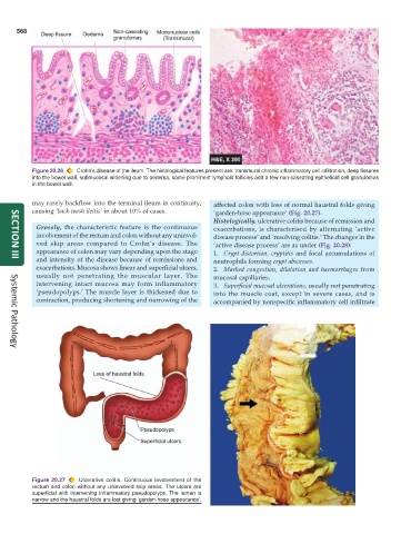
Figure 20.26 Crohn’s disease of the ileum. The histological features present are: transmural chronic inflammatory cell infiltration, deep fissures
into the bowel wall, submucosal widening due to oedema, some prominent lymphoid follicles and a few non-caseating epithelioid cell granulomas
in the bowel wall.
may rarely backflow into the terminal ileum in continuity, affected colon with loss of normal haustral folds giving
causing ‘back-wash ileitis’ in about 10% of cases. ‘garden-hose appearance’ (Fig. 20.27).
Histologically, ulcerative colitis because of remission and
Grossly, the characteristic feature is the continuous exacerbations, is characterised by alternating ‘active
involvement of the rectum and colon without any uninvol- disease process’ and ‘resolving colitis.’ The changes in the
ved skip areas compared to Crohn’s disease. The ‘active disease process’ are as under (Fig. 20.28):
appearance of colon may vary depending upon the stage 1. Crypt distortion, cryptitis and focal accumulations of
and intensity of the disease because of remissions and neutrophils forming crypt abscesses.
SECTION III
exacerbations. Mucosa shows linear and superficial ulcers, 2. Marked congestion, dilatation and haemorrhages from
usually not penetrating the muscular layer. The mucosal capillaries.
intervening intact mucosa may form inflammatory 3. Superficial mucosal ulcerations, usually not penetrating
‘pseudopolyps.’ The muscle layer is thickened due to into the muscle coat, except in severe cases, and is
contraction, producing shortening and narrowing of the accompanied by nonspecific inflammatory cell infiltrate
Systemic Pathology
Figure 20.27 Ulcerative colitis. Continuous involvement of the
rectum and colon without any uninvolved skip areas. The ulcers are
superficial with intervening inflammatory pseudopolyps. The lumen is
narrow and the haustral folds are lost giving ‘garden-hose appearance’.