
Page 585 - Textbook of Pathology, 6th Edition
P. 585
569
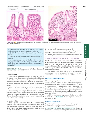
Figure 20.28 Ulcerative colitis in active phase. The microscopic features seen are superficial ulcerations, with mucosal infiltration by inflammatory
cells and a ‘crypt abscess’.
of lymphocytes, plasma cells, neutrophils, some 2. Perianal fistula formation may occur rarely.
eosinophils and mast cells in the lamina propria. 3. Carcinoma may develop in long-standing cases of CHAPTER 20
4. Goblet cells are markedly diminished in cases of active ulcerative colitis of more than 10 years duration.
disease. 4. Stricture formation almost never occurs in ulcerative colitis.
5. Areas of mucosal regeneration and mucodepletion of lin-
ing cells. OTHER INFLAMMATORY LESIONS OF THE BOWEL
6. In long-standing cases, epithelial cytologic atypia Besides IBD, a variety of other acute and chronic inflam-
ranging from mild to marked dysplasia and sometimes matory conditions affect small bowel (enteritis), large bowel
developing into carcinoma in situ and frank adeno- (colitis), or both (enterocolitis); the last named being more
carcinoma.
common. Hence, all these conditions involving small bowel
and/or large bowel are described together here for better
COMPLICATIONS. Complications of Crohn’s disease and correlation of features.
ulcerative colitis are as under: The various forms of inflammations of the bowel after The Gastrointestinal Tract
excluding IBD can be categorised broadly into ‘infective
Crohn’s disease: enterocolitis’ and ‘pseudomembranous enterocolitis.’
1. Malabsorption due to impaired absorption of fat, vitamin
B , proteins and electrolytes from the diseased small bowel. INFECTIVE ENTEROCOLITIS
12
2. Fistula formation may occur in long-standing cases. These
may be internal fistulae between the loops of the intestine, These are a group of acute and chronic inflammatory lesions
or external fistulae such as enterocutaneous, rectal and anal of small intestine and/or colon caused by microorganisms
fistulae. (bacteria, viruses, fungi, protozoa and helminths). All these
3. Stricture formation may occur in chronic cases due to are characterised by diarrhoeal syndromes. Pathogenetically
speaking, these microorganisms can cause enterocolitis by 2
extensive fibrosis in the affected bowel wall. mechanisms—by enteroinvasive bacteria producing ulcerative
4. Development of malignancy in the small intestine as a late lesions, and by enterotoxin-producing bacteria resulting in non-
complication of Crohn’s disease is rarer than that in ulcerative ulcerative lesions.
colitis, but lymphoma may develop more often in Crohn’s A list of common microorganisms producing enterocolitis
disease than adenocarcinoma (seen in some long-standing is presented in Table 20.7. A few important forms are
cases of ulcerative colitis).
described below.
Ulcerative colitis: Intestinal Tuberculosis
1. Toxic megacolon (Fulminant colitis) is the acute fulminating
colitis in which the affected colon is thin-walled and dilated Intestinal tuberculosis can occur in 3 forms—primary,
and is prone to perforation and faecal peritonitis. There is secondary and hyperplastic caecal tuberculosis.
deep penetration of the inflammatory cell infiltrate into 1. PRIMARY INTESTINAL TUBERCULOSIS. Though an
muscle layer which is disrupted. uncommon disease in the developed countries of the world,