
Page 586 - Textbook of Pathology, 6th Edition
P. 586
570
TABLE 20.7: Micro-organisms Causing Infective Microscopically, in the initial stage, there is primary
Enterocolitis. complex or Ghon’s focus in the intestinal mucosa as occurs
A. BACTERIAL ENTEROCOLITIS elsewhere in primary tuberculous infection (page 153).
Subsequently, the mesenteric lymph nodes are affected
1. Entero-invasive bacteria which show typical tuberculous granulomatous
(i) Tuberculosis inflammatory reaction with caseation necrosis.
(ii) Salmonella Tuberculous peritonitis may occur due to spread of the
(iii) Campylobacter jejuni infection.
(iv) Shigella
(v) Escherichia coli 2. SECONDARY INTESTINAL TUBERCULOSIS. Self-
(vi) Yersinia enterocolitica
swallowing of sputum in patients with active pulmonary
2. Enterotoxin-producing bacteria tuberculosis may cause secondary intestinal tuberculosis,
(i) Vibrio cholerae most commonly in the terminal ileum and rarely in the colon.
B. VIRAL ENTEROCOLITIS
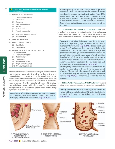
Grossly, the intestinal lesions are prominent than the
C. FUNGAL ENTEROCOLITIS
lesions in regional lymph nodes as in secondary
(i) Candidiasis pulmonary tuberculosis (Fig. 20.29,B). The lesions begin
(ii) Mucormycosis
in the Peyer’s patches or the lymphoid follicles with
D. PROTOZOAL AND METAZOAL INFESTATIONS formation of small ulcers that spread through the
(i) Giardia lamblia lymphatics to form large ulcers which are transverse to the
(ii) Entamoeba histolytica long axis of the bowel, (c.f. typhoid ulcers of small intestine,
(iii) Balantidium coli described below). These ulcers may be coated with caseous
(iv) Taenia solium material. Serosa may be studded with visible tubercles.
(v) Ascaris lumbricoides In advanced cases, transverse fibrous strictures and
(vi) Ancylostoma duodenale intestinal obstruction are seen (Fig. 20.30,A, B).
(vii) Strongyloides stercoralis Histologically, the tuberculous lesions in the intestine are
similar to those observed elsewhere i.e. presence of
primary tuberculosis of the ileocaecal region is quite common tubercles. Mucosa and submucosa show ulceration and
SECTION III
in developing countries including India. In the pre- the muscularis may be replaced by variable degree of
pasteurisation era, it used to occur by ingestion of unpas- fibrosis (Fig. 20.30,C). Tuberculous peritonitis may be
teurised cow’s milk infected with Mycobacterium bovis. But observed.
now-a-days due to control of tuberculosis in cattle and
pasteurisation of milk, virtually all cases of intestinal 3. HYPERPLASTIC CAECAL TUBERCULOSIS. This is a
tuberculosis are caused by M. tuberculosis. The predominant variant of occurring secondary to pulmonary tuberculosis.
changes are in the mesenteric lymph nodes without any
significant intestinal lesion. Grossly, the caecum and/or ascending colon are thick-
walled with mucosal ulceration. Clinically, the lesion is
Grossly, the affected lymph nodes are enlarged, matted palpable and may be mistaken for carcinoma
and caseous (tabes mesenterica). Eventually, there is (Fig. 20.29,C).
healing by fibrosis and calcification (Fig. 20.29,A).
Systemic Pathology
Figure 20.29 Intestinal tuberculosis, three patterns.