
Page 587 - Textbook of Pathology, 6th Edition
P. 587
571
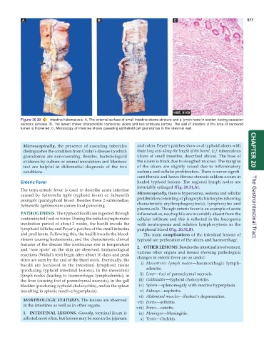
Figure 20.30 Intestinal tuberculosis. A, The external surface of small intestine shows stricture and a lymph node in section having caseation
necrosis (arrows). B, The lumen shows characteristic transverse ulcers and two strictures (arrow). The wall of intestine in the area of narrowed
lumen is thickened. C, Microscopy of intestine shows caseating epithelioid cell granulomas in the intestinal wall.
Microscopically, the presence of caseating tubercles and colon. Peyer’s patches show oval typhoid ulcers with
distinguishes the condition from Crohn’s disease in which their long axis along the length of the bowel, (c.f. tuberculous CHAPTER 20
granulomas are non-caseating. Besides, bacteriological ulcers of small intestine, described above). The base of
evidence by culture or animal inoculation and Mantoux the ulcers is black due to sloughed mucosa. The margins
test are helpful in differential diagnosis of the two of the ulcers are slightly raised due to inflammatory
conditions. oedema and cellular proliferation. There is never signifi-
cant fibrosis and hence fibrous stenosis seldom occurs in
Enteric Fever healed typhoid lesions. The regional lymph nodes are
invariably enlarged (Fig. 20.31,A).
The term enteric fever is used to describe acute infection Microscopically, there is hyperaemia, oedema and cellular
caused by Salmonella typhi (typhoid fever) or Salmonella proliferation consisting of phagocytic histiocytes (showing
paratyphi (paratyphoid fever). Besides these 2 salmonellae,
Salmonella typhimurium causes food poisoning. characteristic erythrophagocytosis), lymphocytes and
plasma cells. Though enteric fever is an example of acute The Gastrointestinal Tract
PATHOGENESIS. The typhoid bacilli are ingested through inflammation, neutrophils are invariably absent from the
contaminated food or water. During the initial asymptomatic cellular infiltrate and this is reflected in the leucopenia
incubation period of about 2 weeks, the bacilli invade the with neutropenia and relative lymphocytosis in the
lymphoid follicles and Peyer’s patches of the small intestine peripheral blood (Fig. 20.31,B).
and proliferate. Following this, the bacilli invade the blood- The main complications of the intestinal lesions of
stream causing bacteraemia, and the characteristic clinical typhoid are perforation of the ulcers and haemorrhage.
features of the disease like continuous rise in temperature 2. OTHER LESIONS. Besides the intestinal involvement,
and ‘rose spots’ on the skin are observed. Immunological various other organs and tissues showing pathological
reactions (Widal’s test) begin after about 10 days and peak changes in enteric fever are as under:
titres are seen by the end of the third week. Eventually, the
bacilli are localised in the intestinal lymphoid tissue i) Mesenteric lymph nodes—haemorrhagic lymph-
(producing typhoid intestinal lesions), in the mesenteric adenitis.
lymph nodes (leading to haemorrhagic lymphadenitis), in ii) Liver—foci of parenchymal necrosis.
the liver (causing foci of parenchymal necrosis), in the gall iii) Gallbladder—typhoid cholecystitis.
bladder (producing typhoid cholecystitis), and in the spleen iv) Spleen—splenomegaly with reactive hyperplasia.
(resulting in splenic reactive hyperplasia). v) Kidneys—nephritis.
vi) Abdominal muscles—Zenker’s degeneration.
MORPHOLOGIC FEATURES. The lesions are observed vii) Joints—arthritis.
in the intestines as well as in other organs.
viii) Bones—osteitis.
1. INTESTINAL LESIONS. Grossly, terminal ileum is ix) Meninges—Meningitis.
affected most often, but lesions may be seen in the jejunum x) Testis—Orchitis.