
Page 588 - Textbook of Pathology, 6th Edition
P. 588
572 described below. Staphylococcal food poisoning occurs due
to liberation of enterotoxins by the bacteria.
2. Clostridial food poisoning. Infection with anaerobic
organisms Clostridium welchii, following consumption of
contaminated meat results in acute food poisoning (page 181).
The illness occurs both by bacterial invasion as well as by
toxins.
3. Botulism. This is a severe form of paralysing illness
caused by ingestion of organism, Clostridium botulinum,
which produces neurotoxin.
4. Salmonella food poisoning (Salmonellosis). This is an
infection (and not caused by toxins) occurring due to food
contaminated by S. typhimurium or S. enteritidis. The condition
manifests with fever, vomiting, and diarrhoea. Death may
result from depletion of water and electrolytes.
Dysenteries
The term ‘dysentery’ is used to mean diarrhoea with
abdominal cramps, tenesmus and passage of mucus in the
stools, from any cause. There are 2 main forms of dysen-
teries—bacillary and amoebic.
1. BACILLARY DYSENTERY. Bacillary dysentery is the
term used for infection by shigella species: S. dysenteriae, S.
flexneri, S. boydii and S. sonnei. Infection occurs by foeco-oral
route and is seen with poor personal hygiene, in densely
populated areas, and with contaminated food and water. The
common housefly plays a role in spread of infection.
Grossly, the lesions are mainly found in the colon and
SECTION III
occasionally in the ileum. Superficial transverse
ulcerations of mucosa of the bowel wall occur in the region
of lymphoid follicles but perforation is seldom seen. The
intervening intact mucosa is hyperaemic and oedematous.
Following recovery from the acute attack, complete
healing usually takes place.
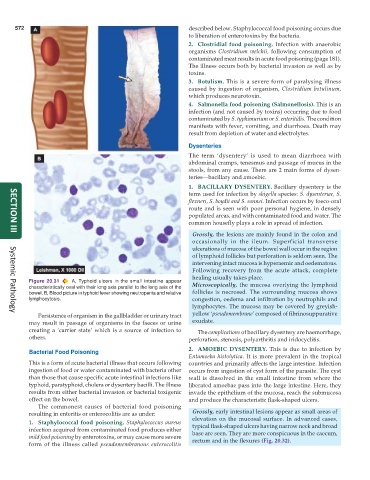
Figure 20.31 A, Typhoid ulcers in the small intestine appear Microscopically, the mucosa overlying the lymphoid
characteristically oval with their long axis parallel to the long axis of the
bowel. B, Blood picture in typhoid fever showing neutropenia and relative follicles is necrosed. The surrounding mucosa shows
lymphocytosis. congestion, oedema and infiltration by neutrophils and
lymphocytes. The mucosa may be covered by greyish-
Persistence of organism in the gallbladder or urinary tract yellow ‘pseudomembrane’ composed of fibrinosuppurative
Systemic Pathology
may result in passage of organisms in the faeces or urine exudate.
creating a ‘carrier state’ which is a source of infection to The complications of bacillary dysentery are haemorrhage,
others. perforation, stenosis, polyarthritis and iridocyclitis.
2. AMOEBIC DYSENTERY. This is due to infection by
Bacterial Food Poisoning
Entamoeba histolytica. It is more prevalent in the tropical
This is a form of acute bacterial illness that occurs following countries and primarily affects the large intestine. Infection
ingestion of food or water contaminated with bacteria other occurs from ingestion of cyst form of the parasite. The cyst
than those that cause specific acute intestinal infections like wall is dissolved in the small intestine from where the
typhoid, paratyphoid, cholera or dysentery bacilli. The illness liberated amoebae pass into the large intestine. Here, they
results from either bacterial invasion or bacterial toxigenic invade the epithelium of the mucosa, reach the submucosa
effect on the bowel. and produce the characteristic flask-shaped ulcers.
The commonest causes of bacterial food poisoning
resulting in enteritis or enterocolitis are as under: Grossly, early intestinal lesions appear as small areas of
elevation on the mucosal surface. In advanced cases,
1. Staphylococcal food poisoning. Staphylococcus aureus typical flask-shaped ulcers having narrow neck and broad
infection acquired from contaminated food produces either base are seen. They are more conspicuous in the caecum,
mild food poisoning by enterotoxins, or may cause more severe rectum and in the flexures (Fig. 20.32).
form of the illness called pseudomembranous enterocolitis