
Page 589 - Textbook of Pathology, 6th Edition
P. 589
573
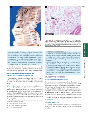
Figure 20.32 Amoebic ulcers large intestine. A, The luminal surface
shows multiple ulcers some of which are deep and are flask-shaped
with narrow neck and broad base (arrow) containing necrotic tissue and
undermined margins. B, Trophozoites of Entamoeba histolytica are seen
at the margin of ulcer (arrow).
Microscopically, the ulcerated area shows chronic MORPHOLOGIC FEATURES. Grossly, the lesions may CHAPTER 20
inflammatory reaction consisting of lymphocytes, plasma be confined, to the large intestine or small intestine, or
cells, macrophages and eosinophils. The trophozoites of both may be involved. The mucosa of the bowel is covered
Entamoeba are seen in the inflammatory exudate and are by patchy, raised yellow-white plaques. Elsewhere, the
concentrated at the advancing margin of the lesion. mucosa is congested and may show small mucosal
Intestinal amoebae characteristically have ingested red ulcerations.
cells in their cytoplasm. Oedema and vascular congestion Microscopically, the ‘pseudomembrane’ is composed of
are present in the area surrounding the ulcers. network of fibrin and mucus, in which are entangled
inflammatory cells and mucosal epithelial cells. There is
Complications of intestinal amoebic ulcers are: amoebic focal necrosis of surface epithelial cells. The lamina propria
liver abscess or amoebic hepatitis, perforation, haemorrhage contains inflammatory cell infiltrate, mainly neutrophils. The Gastrointestinal Tract
and formation of amoeboma which is a tumour-like mass. The submucosa has congested capillaries and may show
microthrombi. The inflammation spreads laterally rather
PSEUDOMEMBRANOUS ENTEROCOLITIS than deeply.
(ANTIBIOTIC-ASSOCIATED DIARRHOEA)
MALABSORPTION SYNDROME
Pseudomembranous enterocolitis is a form of acute
inflammation of colon and/or small intestine characterised DEFINITION AND CLASSIFICATION
by formation of ‘pseudomembrane’ over the site of mucosal The malabsorption syndrome (MAS) is characterised by
injury. impaired intestinal absorption of nutrients especially of fat;
ETIOLOGY. Numerous studies have established the some other substances are proteins, carbohydrates, vitamins
overgrowth of Clostridium difficile with production of its toxin and minerals. MAS is subdivided into 2 broad groups:
in the etiology of antibiotic-associated diarrhoea culminating Primary MAS, which is due to primary deficiency of the
in pseudomembranous colitis. Oral antibiotics such as absorptive mucosal surface and of the associated enzymes.
clindamycin, ampicillin and the cephalosporins are more Secondary MAS, in which mucosal changes result
often (20%) associated with antibiotic-associated diarrhoea, secondary to other factors such as diseases, surgery, trauma
while development of pseudomembranous colitis may occur and drugs.
in 1-10% cases. Each of the two main groups has a number of causes listed
Pseudomembrane formation may also occur in various in Table 20.8.
other conditions as under: CLINICAL FEATURES
Staphylococcal enterocolitis The clinical manifestations of MAS vary according to the
Bacillary (Shigella) dysentery underlying cause. However, some common symptoms are
Candida enterocolitis as follows: