
Page 590 - Textbook of Pathology, 6th Edition
P. 590
574 TABLE 20.8: Classification of Malabsorption Syndrome. INVESTIGATIONS
I. PRIMARY MALABSORPTION When MAS is suspected on clinical grounds, the following
1. Coeliac sprue investigations (laboratory tests) and endoscopic biopsy) may
2. Collagenous sprue be carried out to confirm it:
3. Tropical sprue I. LABORATORY TESTS:
4. Whipple’s disease
5. Disaccharidase deficiency 1. Tests for fat malabsorption:
6. Allergic and eosinophilic gastroenteritis i) Faecal analysis for fat content
ii) Microscopic analysis for faecal fat
II. SECONDARY MALABSORPTION iii) Blood lipid levels after a fatty meal
1. Impaired digestion iv) Tests based on absorption of radioactive-labelled fat.
(i) Mucosal damage e.g. in tuberculosis, Crohn’s disease, lymphoma,
amyloidosis, radiation injury, systemic sclerosis 2. Tests for protein malabsorption:
(ii) Hepatic and pancreatic insufficiency i) Bile acid malabsorption
(iii) Resection of bowel ii) Radioactive-labelled glycine breath test.
(iv) Drugs e.g. methotrexate, neomycin, phenindione etc. iii) Prothrombin time (vitamin K deficiency)
iv) Secretin and other pancreatic tests.
2. Impaired absorption
(i) Short or stagnant bowel (blind loop syndrome) from surgery or 3. Tests for carbohydrate malabsorption:
disease resulting in abnormal proliferation of microbial flora i) D-xylose tolerance test
(ii) Acute infectious enteritis ii) Lactose tolerance test
(iii) Parasitoses e.g. Giardia, Strongyloides, hookworms iii) Hydrogen breath test
iv) Bile acid breath test
3. Impaired transport
(i) Lymphatic obstruction e.g. in lymphoma tuberculosis, lymph- 4. Vitamin B , malabsorption:
12
angiectasia i) Schilling test (page 308).
(ii) Abetalipoproteinaemia
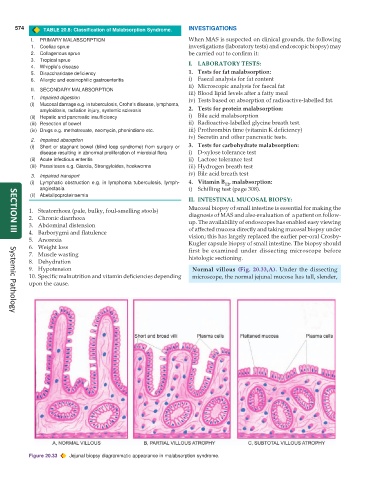
II. INTESTINAL MUCOSAL BIOPSY:
Mucosal biopsy of small intestine is essential for making the
1. Steatorrhoea (pale, bulky, foul-smelling stools)
2. Chronic diarrhoea diagnosis of MAS and also evaluation of a patient on follow-
3. Abdominal distension up. The availability of endoscopes has enabled easy viewing
of affected mucosa directly and taking mucosal biopsy under
4. Barborygmi and flatulence vision; this has largely replaced the earlier per-oral Crosby-
SECTION III
5. Anorexia Kugler capsule biopsy of small intestine. The biopsy should
6. Weight loss first be examined under dissecting microscope before
7. Muscle wasting histologic sectioning.
8. Dehydration
9. Hypotension Normal villous (Fig. 20.33,A). Under the dissecting
10. Specific malnutrition and vitamin deficiencies depending microscope, the normal jejunal mucosa has tall, slender,
upon the cause.
Systemic Pathology
Figure 20.33 Jejunal biopsy diagrammatic appearance in malabsorption syndrome.