
Page 591 - Textbook of Pathology, 6th Edition
P. 591
finger-shaped or leaf-shaped villi. It is lined by tall colum- 575
nar absorptive epithelium and has scattered lymphocytes
in the lamina propria.
Villous atrophy. Variable degree of flattening of intestinal
mucosa in MAS is the commonest pathological change in
mucosal pattern and is referred to as villous atrophy. It
may be of 2 types—partial and subtotal/total type.
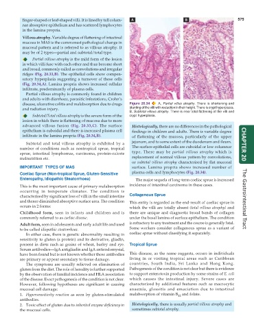
Partial villous atrophy is the mild form of the lesion
in which villi fuse with each other and thus become short
and broad, commonly called as convolutions and irregular
ridges (Fig. 20.33,B). The epithelial cells show compen-
satory hyperplasia suggesting a turnover of these cells
(Fig. 20.34,A). Lamina propria shows increased cellular
infiltrate, predominantly of plasma cells.
Partial villous atrophy is commonly found in children
and adults with diarrhoea, parasitic infestations, Crohn’s
disease, ulcerative colitis and malabsorption due to drugs Figure 20.34 A, Partial villus atrophy. There is shortening and
and radiation injury. blunting of the villi with reduction in their height. There is crypt hyperplasia.
B, Subtotal villous atrophy. There is near total flattening of the villi and
Subtotal/Total villous atrophy is the severe form of the crypt hyperplasia.
lesion in which there is flattening of mucosa due to more
advanced villous fusion (Fig. 20.33,C). The surface Histologically, there are no differences in the pathological
epithelium is cuboidal and there is increased plasma cell findings in children and adults. There is variable degree
infiltrate in the lamina propria (Fig. 20.34,B). of flattening of the mucosa, particularly of the upper
Subtotal and total villous atrophy is exhibited by a jejunum, and to some extent of the duodenum and ileum. CHAPTER 20
number of conditions such as nontropical sprue, tropical The surface epithelial cells are cuboidal or low columnar
sprue, intestinal lymphomas, carcinoma, protein-calorie type. There may be partial villous atrophy which is
malnutrition etc. replacement of normal villous pattern by convolutions,
or subtotal villous atrophy characterised by flat mucosal
IMPORTANT TYPES OF MAS surface. Lamina propria shows increased number of
Coeliac Sprue (Non-tropical Sprue, Gluten-Sensitive plasma cells and lymphocytes (Fig. 20.34).
Enteropathy, Idiopathic Steatorrhoea) The major sequela of long-term coeliac sprue is increased
This is the most important cause of primary malabsorption incidence of intestinal carcinoma in these cases.
occurring in temperate climates. The condition is
characterised by significant loss of villi in the small intestine Collagenous Sprue
and thence diminished absorptive surface area. The condition This entity is regarded as the end-result of coeliac sprue in The Gastrointestinal Tract
occurs in 2 forms: which the villi are totally absent (total villous atrophy) and
Childhood form, seen in infants and children and is there are unique and diagnostic broad bands of collagen
commonly referred to as coeliac disease. under the basal lamina of surface epithelium. The condition
Adult form, seen in adolescents and early adult life and used is refractory to any treatment and the course is generally fatal.
to be called idiopathic steatorrhoea. Some workers consider collagenous sprue as a variant of
In either case, there is genetic abnormality resulting in coeliac sprue without classifying it separately.
sensitivity to gluten (a protein) and its derivative, gliadin,
present in diets such as grains of wheat, barley and rye. Tropical Sprue
Serum antibodies—IgA antigliadin and IgA antiendomysial,
have been found but is not known whether these antibodies This disease, as the name suggests, occurs in individuals
are primary or appear secondary to tissue damage. living in or visiting tropical areas such as Caribbean
The symptoms are usually relieved on elimination of countries, South India, Sri Lanka and Hong Kong.
gluten from the diet. The role of heredity is further supported Pathogenesis of the condition is not clear but there is evidence
by the observation of familial incidence and HLA association to support enterotoxin production by some strains of E. coli
of the disease. Exact pathogenesis of the condition is not clear. which causes the intestinal injury. Severe cases are
However, following hypotheses are significant in causing characterised by additional features such as macrocytic
mucosal cell damage: anaemia, glossitis and emaciation due to intestinal
1. Hypersensitivity reaction as seen by gluten-stimulated malabsorption of vitamin B and folate.
12
antibodies.
2. Toxic effect of gluten due to inherited enzyme deficiency in Histologically, there is usually partial villous atrophy and
the mucosal cells. sometimes subtotal atrophy.