
Page 592 - Textbook of Pathology, 6th Edition
P. 592
576 The lesions are relieved by removal of the patient from
the tropical area and by oral administration of antibiotics
but gluten-free diet has no role in improvement.
Whipple’s Disease (Intestinal Lipodystrophy)
This is an uncommon bacterial disease involving not only
the intestines but also various other systems such as central
nervous system, heart, blood vessels, skin, joints, lungs, liver,
spleen and kidneys. The disease is more common in males
in 4th to 5th decades of life. Patients may present with
features of malabsorption or may have atypical presentation
in the form of migratory polyarthritis, neurological
disturbances and focal hyperpigmentation of the skin.
Histologically, the affected tissues show presence of
characteristic macrophages containing PAS-positive
granules and rod-shaped micro-organisms (Whipple’s
bacilli). These macrophages are predominantly present in
the lamina propria of the small intestine and mesenteric
lymph nodes.
Patients respond very well to oral antibiotic therapy.
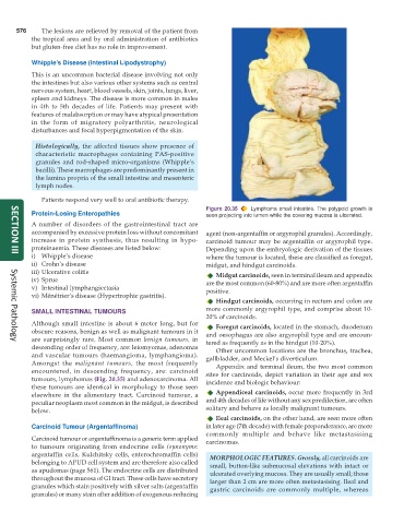
Figure 20.35 Lymphoma small intestine. The polypoid growth is
Protein-Losing Enteropathies seen projecting into lumen while the covering mucosa is ulcerated.
A number of disorders of the gastrointestinal tract are
accompanied by excessive protein loss without concomitant agent (non-argentaffin or argyrophil granules). Accordingly,
increase in protein synthesis, thus resulting in hypo- carcinoid tumour may be argentaffin or argyrophil type.
proteinaemia. These diseases are listed below: Depending upon the embryologic derivation of the tissues
i) Whipple’s disease where the tumour is located, these are classified as foregut,
SECTION III
ii) Crohn’s disease midgut, and hindgut carcinoids.
iii) Ulcerative colitis Midgut carcinoids, seen in terminal ileum and appendix
iv) Sprue are the most common (60-80%) and are more often argentaffin
v) Intestinal lymphangiectasia positive.
vi) Ménétrier’s disease (Hypertrophic gastritis).
Hindgut carcinoids, occurring in rectum and colon are
SMALL INTESTINAL TUMOURS more commonly argyrophil type, and comprise about 10-
20% of carcinoids.
Although small intestine is about 6 meter long, but for Foregut carcinoids, located in the stomach, duodenum
obscure reasons, benign as well as malignant tumours in it and oesophagus are also argyrophil type and are encoun-
are surprisingly rare. Most common benign tumours, in tered as frequently as in the hindgut (10-20%).
Systemic Pathology
descending order of frequency, are: leiomyomas, adenomas Other uncommon locations are the bronchus, trachea,
and vascular tumours (haemangioma, lymphangioma). gallbladder, and Meckel’s diverticulum.
Amongst the malignant tumours, the most frequently Appendix and terminal ileum, the two most common
encountered, in descending frequency, are: carcinoid sites for carcinoids, depict variation in their age and sex
tumours, lymphomas (Fig. 20.35) and adenocarcinoma. All incidence and biologic behaviour:
these tumours are identical in morphology to those seen
elsewhere in the alimentary tract. Carcinoid tumour, a Appendiceal carcinoids, occur more frequently in 3rd
peculiar neoplasm most common in the midgut, is described and 4th decades of life without any sex predilection, are often
below. solitary and behave as locally malignant tumours.
Ileal carcinoids, on the other hand, are seen more often
Carcinoid Tumour (Argentaffinoma) in later age (7th decade) with female preponderance, are more
commonly multiple and behave like metastasising
Carcinoid tumour or argentaffinoma is a generic term applied carcinomas.
to tumours originating from endocrine cells (synonyms:
argentaffin cells, Kulchitsky cells, enterochromaffin cells) MORPHOLOGIC FEATURES. Grossly, all carcinoids are
belonging to APUD cell system and are therefore also called small, button-like submucosal elevations with intact or
as apudomas (page 561). The endocrine cells are distributed ulcerated overlying mucosa. They are usually small; those
throughout the mucosa of GI tract. These cells have secretory larger than 2 cm are more often metastasising. Ileal and
granules which stain positively with silver salts (argentaffin gastric carcinoids are commonly multiple, whereas
granules) or many stain after addition of exogenous reducing