
Page 594 - Textbook of Pathology, 6th Edition
P. 594
578
Histologically, appendix has four layers in its wall—
mucosa, submucosa, muscularis and serosa. The mucosa has
patchy distribution of crypts and the submucosa has
abundant lymphoid tissue. Argentaffin and non-
argentaffin endocrine cells are present in the base of
mucosal glands just as in the small intestine. The muscu-
laris of the appendix has two layers (inner circular and
outer longitudinal) as elsewhere in the alimentary tract.
Two important diseases involving the appendix are
appendicitis and appendiceal carcinoids.
APPENDICITIS
Acute inflammation of the appendix, acute appendicitis, is
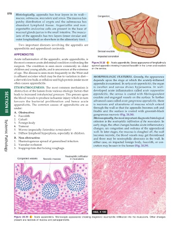
the most common acute abdominal condition confronting the Figure 20.38 Acute appendicitis. Gross appearance of longitudinally
surgeon. The condition is seen more commonly in older opened appendix showing impacted faecolith in the lumen and exudate
children and young adults, and is uncommon at the extremes on the serosa.
of age. The disease is seen more frequently in the West and
in affluent societies which may be due to variation in diet— MORPHOLOGIC FEATURES. Grossly, the appearance
a diet with low bulk or cellulose and high protein intake more depends upon the stage at which the acutely-inflamed
often causes appendicitis. appendix is examined. In early acute appendicitis, the organ
ETIOPATHOGENESIS. The most common mechanism is is swollen and serosa shows hyperaemia. In well-
obstruction of the lumen from various etiologic factors that developed acute inflammation called acute suppurative
leads to increased intraluminal pressure. This presses upon appendicitis, the serosa is coated with fibrinopurulent
the blood vessels to produce ischaemic injury which in turn exudate and engorged vessels on the surface. In further
favours the bacterial proliferation and hence acute advanced cases called acute gangrenous appendicitis, there
appendicitis. The common causes of appendicitis are as is necrosis and ulcerations of mucosa which extend
under: through the wall so that the appendix becomes soft and
A. Obstructive: friable and the surface is coated with greenish-black
SECTION III
1. Faecolith gangrenous necrosis (Fig. 20.38).
2. Calculi Microscopically, the most important diagnostic histological
3. Foreign body criterion is the neutrophilic infiltration of the muscularis. In
4. Tumour early stage, the other changes besides acute inflammatory
changes, are congestion and oedema of the appendiceal
5. Worms (especially Enterobius vermicularis) wall. In later stages, the mucosa is sloughed off, the wall
6. Diffuse lymphoid hyperplasia, especially in children.
becomes necrotic, the blood vessels may get thrombosed
B. Non-obstructive: and there may be neutrophilic abscesses in the wall. In
1. Haematogenous spread of generalised infection either case, an impacted foreign body, faecolith, or con-
2. Vascular occlusion cretion may be seen in the lumen (Fig. 20.39).
3. Inappropriate diet lacking roughage.
Systemic Pathology
Figure 20.39 Acute appendicitis. Microscopic appearance showing diagnostic neutrophilic infiltration into the muscularis. Other changes
present are necrosis of mucosa and periappendicitis.