
Page 596 - Textbook of Pathology, 6th Edition
P. 596
580 CONGENITAL MALFORMATIONS
Hirschsprung’s Disease (Congenital Megacolon)
The term ‘megacolon’ is used for any form of marked
dilatation of the entire colon or its segment and may occur
as a congenital or acquired disorder. Congenital form
characterised by congenital absence of ganglion cells in the
bowel wall (enteric neurons) is called Hirschsprung’s disease.
As a result, the aganglionic segment remains contracted.
Genetically, Hirschsprung’s disease is a heterogeneous
disorder as under:
1. Autosomal dominant inheritance with mutation in RET
proto-oncogene in some cases.
2. Autosomal recessive form with mutation in endothelin-B
receptor gene in many other cases.
Clinically, the condition manifests shortly after birth with
constipation, gaseous distension and sometimes with acute Figure 20.40 Hirschsprung’s disease, diagrammatic representation
intestinal obstruction. Its frequency is 1 in 5,000 live-births, of the pathologic changes.
has familial tendency in about 4% of cases and has
predilection for development in Down’s syndrome. 5. Zonal colonic aganglionosis: A short segment is nvolved in
Pathogenesis lies in the failure of neuroblasts to migrate to agnaglionosis in which the ganglia cells are absent both above
the rectum which normally occurs at about 12 weeks of and below the aganglionic segment.
gestation.
In addition to congenital megacolon discussed above,
megacolon may occur from certain acquired causes as under:
MORPHOLOGIC FEATURES. Two types of biopsies may i. Obstructive e.g. due to tumour, post-inflammatory
be done on infants suspected of having Hirschsprung’s strictures.
disease—full-thickness rectal biopsy, and suction biopsy ii. Endocrine e.g. in myxoedema, cretinism.
that includes mucosa and submucosa. iii. CNS disorders e.g. spina bifida, paraplegia, parkinsonism.
SECTION III
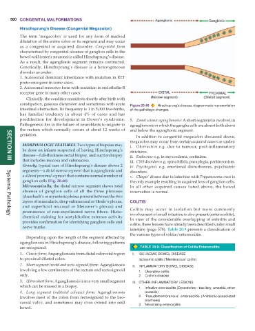
Grossly, typical case of Hirschsprung’s disease shows 2 iv. Psychogenic e.g. emotional disturbances, psychiatric
segments—a distal narrow segment that is aganglionic and disorders.
a dilated proximal segment that contains normal number of v. Chagas’ disease due to infection with Trypanosoma cruzi is
ganglion cells (Fig. 20.40). the only example resulting in acquired loss of ganglion cells.
Microscopically, the distal narrow segment shows total In all other acquired causes listed above, the bowel
absence of ganglion cells of all the three plexuses innervation is normal.
(Auerbach’s or myenteric plexus present between the two
layers of muscularis, deep submucosal or Henle’s plexus, COLITIS
and superficial mucosal or Meissner’s plexus) and
prominence of non-myelinated nerve fibres. Histo- Colitis may occur in isolation but more commonly
involvement of small intestine is also present (enterocolitis).
chemical staining for acetylcholine esterase activity In view of the considerable overlapping of enteritis and
Systemic Pathology
provides confirmation for identifying ganglion cells and colitis, these lesions have already been described under small
nerve trunks.
intestine (page 578). Table 20.9 presents a classification of
the various types of colitis/enterocolitis.
Depending upon the length of the segment affected by
aganglionosis in Hirschsprung’s disease, following patterns
are recognised: TABLE 20.9: Classification of Colitis/Enterocolitis.
1. Classic form: Anganglionosis from distal colorectal region I. ISCHAEMIC BOWEL DISEASE
to proximal dilated colon. Ischaemic colitis (‘Membranous’ colitis)
2. Short segment (rectal and recto-sigmoid) form: Aganglionosis II. INFLAMMATORY BOWEL DISEASE
involving a few centimeters of the rectum and rectosigmoid 1. Ulcerative colitis
only. 2. Crohn’s disease
3. Ultra-short form: Aganglionosis is in a very small segment III. OTHER INFLAMMATORY LESIONS
which can be missed in a biopsy. 1. Infective enterocolitis (Dysenteries—bacillary, amoebic, other
4. Long segment (subtotal colonic) form: Aganglionosis parasitic)
involves most of the colon from rectosigmoid to the ileo- 2. ‘Pseudomembranous’ enterocolitis (Antibiotic-associated
caecal valve, and sometimes may even extend into smll 3. diarrhoea)
Necrotising enterocolitis
bowel.