
Page 600 - Textbook of Pathology, 6th Edition
P. 600
584
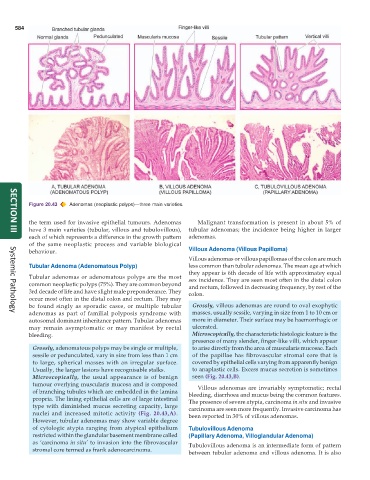
Figure 20.43 Adenomas (neoplastic polyps)—three main varieties.
the term used for invasive epithelial tumours. Adenomas Malignant transformation is present in about 5% of
have 3 main varieties (tubular, villous and tubulovillous), tubular adenomas; the incidence being higher in larger
SECTION III
each of which represents a difference in the growth pattern adenomas.
of the same neoplastic process and variable biological
behaviour. Villous Adenoma (Villous Papilloma)
Villous adenomas or villous papillomas of the colon are much
Tubular Adenoma (Adenomatous Polyp) less common than tubular adenomas. The mean age at which
they appear is 6th decade of life with approximatey equal
Tubular adenomas or adenomatous polyps are the most sex incidence. They are seen most often in the distal colon
common neoplastic polyps (75%). They are common beyond and rectum, followed in decreasing frequency, by rest of the
3rd decade of life and have slight male preponderance. They colon.
occur most often in the distal colon and rectum. They may
be found singly as sporadic cases, or multiple tubular Grossly, villous adenomas are round to oval exophytic
adenomas as part of familial polyposis syndrome with masses, usually sessile, varying in size from 1 to 10 cm or
Systemic Pathology
autosomal dominant inheritance pattern. Tubular adenomas more in diameter. Their surface may be haemorrhagic or
may remain asymptomatic or may manifest by rectal ulcerated.
bleeding. Microscopically, the characteristic histologic feature is the
presence of many slender, finger-like villi, which appear
Grossly, adenomatous polyps may be single or multiple, to arise directly from the area of muscularis mucosae. Each
sessile or pedunculated, vary in size from less than 1 cm of the papillae has fibrovascular stromal core that is
to large, spherical masses with an irregular surface. covered by epithelial cells varying from apparently benign
Usually, the larger lesions have recognisable stalks. to anaplastic cells. Excess mucus secretion is sometimes
Microscopically, the usual appearance is of benign seen (Fig. 20.43,B).
tumour overlying muscularis mucosa and is composed Villous adenomas are invariably symptomatic; rectal
of branching tubules which are embedded in the lamina bleeding, diarrhoea and mucus being the common features.
propria. The lining epithelial cells are of large intestinal The presence of severe atypia, carcinoma in situ and invasive
type with diminished mucus secreting capacity, large carcinoma are seen more frequently. Invasive carcinoma has
nuclei and increased mitotic activity (Fig. 20.43,A). been reported in 30% of villous adenomas.
However, tubular adenomas may show variable degree
of cytologic atypia ranging from atypical epithelium Tubulovillous Adenoma
restricted within the glandular basement membrane called (Papillary Adenoma, Villoglandular Adenoma)
as ‘carcinoma in situ’ to invasion into the fibrovascular Tubulovillous adenoma is an intermediate form of pattern
stromal core termed as frank adenocarcinoma.
between tubular adenoma and villous adenoma. It is also